

Contributions
Abstract: EP866
Type: E-Poster Presentation
Session title: Lymphoma Biology & Translational Research
Background
Diffuse large B cell lymphoma (DLBCL) is the most common non-Hodgkin's lymphoma (NHL) with high heterogeneity. Previous studies have identified that NUSAP1 plays an important role in the occurrence and progress of multiple tumors. Whereas, no research has been reported regarding the role of NUSAP1 in DLBCL.
Aims
Herein, we aim to further explore the expression and exact role of NUSAP1 in DLBCL development, and provide theoretical basis for new molecular markers and targeted therapy.
Methods
Peripheral blood samples from de novo DLBCL patients and healthy volunteers were collected with informed consents at the Department of Hematology in Shandong Provincial Hospital Affiliated to Shandong University (SPHASU). Kaplan-Meier survival curves with log-rank test of overall survival (OS) were analyzed. Immunohistochemistry staining (IHC) was performed to assess NUSAP1 expression in specimens. Expression levels of NUSAP1 mRNA and protein were detected by quantitative RT-PCR and western blotting. The DLBCL cells were transfected by lentiviral shRNA and vectors to stably silence and up-regulate NUSAP1. Effects of doxorubicin on cell viabilities were assessed by cell counting kit-8. Besides, apoptosis and cell cycle were respectively detected by annexin V-PE/7AAD and PI/RNase staining via flow cytometry. Invasion ability was analyzed by transwell assay. ShNUSAP1 cells and Scramble cells were subcutaneously injected to SCID-Beige mice to establish xenograft models.
Results
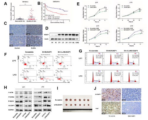
According to clinical specimens and bioinformatics analysis, the expression level of NUSAP1 gene in samples of DLBCL patients was significantly increased than that of healthy donors (P<0.05) (Figure A). Besides, patients with high expression of NUSAP1 was related to shorter overall survival (HR=0.456, P<0.01), indicating poor prognosis (Figure B). Stronger positivity of NUSAP1 was markedly observed in DLBCL lymph nodes than reactive hyperplasia group, which was associated with Ann Arbor stage of DLBCL patients (Figure C). Compared with normal B cells, the protein level of NUSAP1 were up-regulated in DLBCL cell lines (Figure D). After transfected with lentivirus, the proliferation rate of NUSAP1 knockdown group was lower than that of the control group, while the overexpression group was faster than control (Figure E). Through flow cytometry, silencing of NUSAP1 led to increased apoptotic rates of LY1 and LY3 cells (Figure F). Along with the recovery of NUSAP1 expression level, apoptosis rates were improved again. With the addition of doxorubicin at 100nM, interference of NUSAP1 could increase the sensitivity of cells to doxorubicin (Figure E). NUSAP1 intervention induced obvious cell cycle arrest in G1 phase of LY1 and LY3 cell lines, with concomitant reduction of cell proportion in S phase (Figure G). Surppression of NUSAP1 inhibited the action of DNA damage repair pathway (Figure H). We found that tumor growth was significantly suppressed by inhibiting NUSAP1 in xenograft models (Figure I). IHC for NUSAP1 and Ki67 on tumor sections showed that tumors derived from shNUSAP1 cells exhibited significantly lower levels of Ki67 compared to control group (Figure J).
Conclusion
Collectively, NUSAP1 contributes to tumor growth both in vivo and vitro through DNA damage repair pathway, which providing a new direction for prognosis assessment and targeted therapy of DLBCL.
Keyword(s): Diffuse large B cell lymphoma, DNA damage, Oncogene
Abstract: EP866
Type: E-Poster Presentation
Session title: Lymphoma Biology & Translational Research
Background
Diffuse large B cell lymphoma (DLBCL) is the most common non-Hodgkin's lymphoma (NHL) with high heterogeneity. Previous studies have identified that NUSAP1 plays an important role in the occurrence and progress of multiple tumors. Whereas, no research has been reported regarding the role of NUSAP1 in DLBCL.
Aims
Herein, we aim to further explore the expression and exact role of NUSAP1 in DLBCL development, and provide theoretical basis for new molecular markers and targeted therapy.
Methods
Peripheral blood samples from de novo DLBCL patients and healthy volunteers were collected with informed consents at the Department of Hematology in Shandong Provincial Hospital Affiliated to Shandong University (SPHASU). Kaplan-Meier survival curves with log-rank test of overall survival (OS) were analyzed. Immunohistochemistry staining (IHC) was performed to assess NUSAP1 expression in specimens. Expression levels of NUSAP1 mRNA and protein were detected by quantitative RT-PCR and western blotting. The DLBCL cells were transfected by lentiviral shRNA and vectors to stably silence and up-regulate NUSAP1. Effects of doxorubicin on cell viabilities were assessed by cell counting kit-8. Besides, apoptosis and cell cycle were respectively detected by annexin V-PE/7AAD and PI/RNase staining via flow cytometry. Invasion ability was analyzed by transwell assay. ShNUSAP1 cells and Scramble cells were subcutaneously injected to SCID-Beige mice to establish xenograft models.
Results
According to clinical specimens and bioinformatics analysis, the expression level of NUSAP1 gene in samples of DLBCL patients was significantly increased than that of healthy donors (P<0.05) (Figure A). Besides, patients with high expression of NUSAP1 was related to shorter overall survival (HR=0.456, P<0.01), indicating poor prognosis (Figure B). Stronger positivity of NUSAP1 was markedly observed in DLBCL lymph nodes than reactive hyperplasia group, which was associated with Ann Arbor stage of DLBCL patients (Figure C). Compared with normal B cells, the protein level of NUSAP1 were up-regulated in DLBCL cell lines (Figure D). After transfected with lentivirus, the proliferation rate of NUSAP1 knockdown group was lower than that of the control group, while the overexpression group was faster than control (Figure E). Through flow cytometry, silencing of NUSAP1 led to increased apoptotic rates of LY1 and LY3 cells (Figure F). Along with the recovery of NUSAP1 expression level, apoptosis rates were improved again. With the addition of doxorubicin at 100nM, interference of NUSAP1 could increase the sensitivity of cells to doxorubicin (Figure E). NUSAP1 intervention induced obvious cell cycle arrest in G1 phase of LY1 and LY3 cell lines, with concomitant reduction of cell proportion in S phase (Figure G). Surppression of NUSAP1 inhibited the action of DNA damage repair pathway (Figure H). We found that tumor growth was significantly suppressed by inhibiting NUSAP1 in xenograft models (Figure I). IHC for NUSAP1 and Ki67 on tumor sections showed that tumors derived from shNUSAP1 cells exhibited significantly lower levels of Ki67 compared to control group (Figure J).
Conclusion
Collectively, NUSAP1 contributes to tumor growth both in vivo and vitro through DNA damage repair pathway, which providing a new direction for prognosis assessment and targeted therapy of DLBCL.
Keyword(s): Diffuse large B cell lymphoma, DNA damage, Oncogene