

Contributions
Abstract: EP647
Type: E-Poster Presentation
Session title: Chronic lymphocytic leukemia and related disorders - Clinical
Background
The most used methods for assessing minimal residual disease (MRD) in CLL are based on flow cytometry or RQ-PCR with allele-specific oligonucleotide primers. Recently, the assessment of MRD by the level of specific extracellular DNA using the most sensitive ddPCR technologies, as well as NGS approaches, has been proposed. Blood mRNA and microRNA molecules can also be studied as probable informative biomarkers of MRD in CLL (Giudice I.D., et.al., 2019). The use of plasma nucleic acids promises to be more effective in assessing MRD because they can detect the presence of B-CLL cells in different compartments, including lymph nodes and bone marrow. A new flow cytometric method for assessing MRD status has been proposed based on analysis of the tumor-associated antigen ROR1 (CD160-ROR1 FCA) (Farren T.W., et. al., 2020). ROR1, a receptor transmembrane tyrosine kinase, is highly expressed by cells during embryonic development but is not found in adult tissues. Expression of antigen ROR1 has been found in patients with B-CLL cells and independent of anatomical niches, clinical B-CLL heterogeneity, or B-cell activation. Another knowledge onco-associated RNA in CLL is mir-155 (Stolyar M.A., et. al., 2020; Cui B., et. al., 2014).
Aims
To investigate the usefulness of the combination of two ROR1 mRNA and miR-155 for evaluating the effectiveness of CLL therapy.
Methods
This study included blood samples from 37 B-CLL patients and 9 of it were observed before and during therapy of fludarabine, cyclophosphamide, rituximab combination. The control group consisted of 51 healthy donors. The expression level of ROR1 mRNA and miR-155 was determined by RT-PCR using Formula Gena Ltd kits. The results were statistically evaluated using the R- package.
Results
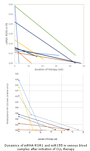
We did not find expression of mRNA ROR1 in venous blood and miR-155 in plasma of healthy donors. Overexpression of ROR1 (Me=0.16) was observed in 70% of our CLL patients before treatment. miR-155 level in this group was Me=0.05 relative units. Both RNAs were weakly dependent on the blood cells count or disease stage. Initially, increased ROR1 expression decreased in all patients during therapy (Figure). In most patients, the level of circulating miRNA-155 in the blood plasma also decreased (Figure). RNA dynamics correlated with clinical improvement. Further enhancement the analytical sensitivity of PCR for selected RNAs may be useful in developing new approaches to assessing MRD in CLL. It is important to note that for these tests there is no need for preliminary sequencing of allele-specific regions of immunoglobulin genes, as well as in expensive devices for NGS.
Conclusion
The dynamics of mRNA ROR1 and miR-155 levels could be explored in the future as biomarkers of residual disease in CLL
Keyword(s): Chronic lymphocytic leukemia, RT-PCR
Abstract: EP647
Type: E-Poster Presentation
Session title: Chronic lymphocytic leukemia and related disorders - Clinical
Background
The most used methods for assessing minimal residual disease (MRD) in CLL are based on flow cytometry or RQ-PCR with allele-specific oligonucleotide primers. Recently, the assessment of MRD by the level of specific extracellular DNA using the most sensitive ddPCR technologies, as well as NGS approaches, has been proposed. Blood mRNA and microRNA molecules can also be studied as probable informative biomarkers of MRD in CLL (Giudice I.D., et.al., 2019). The use of plasma nucleic acids promises to be more effective in assessing MRD because they can detect the presence of B-CLL cells in different compartments, including lymph nodes and bone marrow. A new flow cytometric method for assessing MRD status has been proposed based on analysis of the tumor-associated antigen ROR1 (CD160-ROR1 FCA) (Farren T.W., et. al., 2020). ROR1, a receptor transmembrane tyrosine kinase, is highly expressed by cells during embryonic development but is not found in adult tissues. Expression of antigen ROR1 has been found in patients with B-CLL cells and independent of anatomical niches, clinical B-CLL heterogeneity, or B-cell activation. Another knowledge onco-associated RNA in CLL is mir-155 (Stolyar M.A., et. al., 2020; Cui B., et. al., 2014).
Aims
To investigate the usefulness of the combination of two ROR1 mRNA and miR-155 for evaluating the effectiveness of CLL therapy.
Methods
This study included blood samples from 37 B-CLL patients and 9 of it were observed before and during therapy of fludarabine, cyclophosphamide, rituximab combination. The control group consisted of 51 healthy donors. The expression level of ROR1 mRNA and miR-155 was determined by RT-PCR using Formula Gena Ltd kits. The results were statistically evaluated using the R- package.
Results
We did not find expression of mRNA ROR1 in venous blood and miR-155 in plasma of healthy donors. Overexpression of ROR1 (Me=0.16) was observed in 70% of our CLL patients before treatment. miR-155 level in this group was Me=0.05 relative units. Both RNAs were weakly dependent on the blood cells count or disease stage. Initially, increased ROR1 expression decreased in all patients during therapy (Figure). In most patients, the level of circulating miRNA-155 in the blood plasma also decreased (Figure). RNA dynamics correlated with clinical improvement. Further enhancement the analytical sensitivity of PCR for selected RNAs may be useful in developing new approaches to assessing MRD in CLL. It is important to note that for these tests there is no need for preliminary sequencing of allele-specific regions of immunoglobulin genes, as well as in expensive devices for NGS.
Conclusion
The dynamics of mRNA ROR1 and miR-155 levels could be explored in the future as biomarkers of residual disease in CLL
Keyword(s): Chronic lymphocytic leukemia, RT-PCR