

Contributions
Abstract: EP605
Type: E-Poster Presentation
Session title: Chronic lymphocytic leukemia and related disorders - Biology & Translational Research
Background
Enhanced DNA damage repair effect is an important mechanism for drug-resistance in chronic lymphocytic leukemia (CLL). Previous studies reported that nucleolar and spindle associated protein 1 (NUSAP1) was involved in DNA damage repair process and played important roles in the development, progression, and metastasis in several types of cancer. However, its role and mechanism in the development of CLL are still unclear.
Aims
We aim to further explore the expression and exact role of NUSAP1 in CLL development, providing theoretical basis for new molecular markers and targeted therapy.
Methods
Expression levels of NUSAP1 mRNA and protein in CLL cell lines and patient specimens were detected by qRT-PCR and Western blot, and Kaplan-Meier survival curve and overall survival were analyzed by log-rank test. Peripheral blood samples from de novo CLL patients and healthy volunteers were collected with informed consents at the Department of Hematology in Shandong Provincial Hospital Affiliated to Shandong University (SPHASU). Microarray datasets GSE22762 were obtained from Gene Expression Omnibus. With altering NUSAP1 expression by lentivirus-transfected cells in vitro, the effects of NUSAP1 on cell proliferation, apoptosis and cycle were detected by CCK8, Annexin V-PE /7AAD staining and PI/RNase staining respectively.
Results
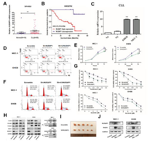
To study the role of NUSAP1, CLL specimens were chosen for qRT-PCR analysis. The expression level of NUSAP1 gene in samples of CLL patients was significantly increased than that of healthy donors (P<0.05) (Figure A). Besides, the results indicated that the overall survival (OS) of patients with highly expressed NUSAP1 was significantly worse than in patients with low expression with the statistical analysis database GSE22762 (Figure B). mRNA and protein expression levels of NUSAP1 were significantly higher in CLL cell lines than in PBMCs from healthy donors (Figure C). Moreover, the amounts of DNA fragmentation of the apoptotic cells were remarkably increased in shNUSAP1 transfection cells compared with the Scramble group (Figure D). Our findings indicated that NUSAP1 knockdown notably suppressed cell proliferation (Figure E). In addition, after downregulating NUSAP1, MEC-1 and EHEB cells were blocked in G0/G1 phase (Figure F). Treatment with fludarabine or ibrutinib in shNUSAP1 group showed enhanced cytotoxicity in CLL cells (Figure G). Suppression of NUSAP1 inhibited the action of proteins in DNA damage repair pathway (Figure H). Through COIP, NUSAP1 was identified to bind with RAD51 (Figure H). After CLL cells were subcutaneously injected into (SCID) beige mice for 45 days, tumor volumes were significantly reduced in mice treated with ShNUSAP1 cells compared to scramble group (Figure I). Compared with the NUSAP1 upregulated group, the NUSAP1-ΔSAP transfection group suppressed RAD51 expression as expected (Figure J). Hence, NUSAP1 participates in the DNA damage repair process and enhances the drug resistance in CLL.
Conclusion
Collectively, our findings demonstrate that NUSAP1 contributes to DNA damage repairing by binding with RAD51 and promoting stable proliferation of CLL cells both in vitro and vivo. Therefore, NUSAP1 is expected to be a potential target for the treatment of CLL with drug-resistance.
Keyword(s): DNA damage, Oncogene, Prognosis
Abstract: EP605
Type: E-Poster Presentation
Session title: Chronic lymphocytic leukemia and related disorders - Biology & Translational Research
Background
Enhanced DNA damage repair effect is an important mechanism for drug-resistance in chronic lymphocytic leukemia (CLL). Previous studies reported that nucleolar and spindle associated protein 1 (NUSAP1) was involved in DNA damage repair process and played important roles in the development, progression, and metastasis in several types of cancer. However, its role and mechanism in the development of CLL are still unclear.
Aims
We aim to further explore the expression and exact role of NUSAP1 in CLL development, providing theoretical basis for new molecular markers and targeted therapy.
Methods
Expression levels of NUSAP1 mRNA and protein in CLL cell lines and patient specimens were detected by qRT-PCR and Western blot, and Kaplan-Meier survival curve and overall survival were analyzed by log-rank test. Peripheral blood samples from de novo CLL patients and healthy volunteers were collected with informed consents at the Department of Hematology in Shandong Provincial Hospital Affiliated to Shandong University (SPHASU). Microarray datasets GSE22762 were obtained from Gene Expression Omnibus. With altering NUSAP1 expression by lentivirus-transfected cells in vitro, the effects of NUSAP1 on cell proliferation, apoptosis and cycle were detected by CCK8, Annexin V-PE /7AAD staining and PI/RNase staining respectively.
Results
To study the role of NUSAP1, CLL specimens were chosen for qRT-PCR analysis. The expression level of NUSAP1 gene in samples of CLL patients was significantly increased than that of healthy donors (P<0.05) (Figure A). Besides, the results indicated that the overall survival (OS) of patients with highly expressed NUSAP1 was significantly worse than in patients with low expression with the statistical analysis database GSE22762 (Figure B). mRNA and protein expression levels of NUSAP1 were significantly higher in CLL cell lines than in PBMCs from healthy donors (Figure C). Moreover, the amounts of DNA fragmentation of the apoptotic cells were remarkably increased in shNUSAP1 transfection cells compared with the Scramble group (Figure D). Our findings indicated that NUSAP1 knockdown notably suppressed cell proliferation (Figure E). In addition, after downregulating NUSAP1, MEC-1 and EHEB cells were blocked in G0/G1 phase (Figure F). Treatment with fludarabine or ibrutinib in shNUSAP1 group showed enhanced cytotoxicity in CLL cells (Figure G). Suppression of NUSAP1 inhibited the action of proteins in DNA damage repair pathway (Figure H). Through COIP, NUSAP1 was identified to bind with RAD51 (Figure H). After CLL cells were subcutaneously injected into (SCID) beige mice for 45 days, tumor volumes were significantly reduced in mice treated with ShNUSAP1 cells compared to scramble group (Figure I). Compared with the NUSAP1 upregulated group, the NUSAP1-ΔSAP transfection group suppressed RAD51 expression as expected (Figure J). Hence, NUSAP1 participates in the DNA damage repair process and enhances the drug resistance in CLL.
Conclusion
Collectively, our findings demonstrate that NUSAP1 contributes to DNA damage repairing by binding with RAD51 and promoting stable proliferation of CLL cells both in vitro and vivo. Therefore, NUSAP1 is expected to be a potential target for the treatment of CLL with drug-resistance.
Keyword(s): DNA damage, Oncogene, Prognosis