

Contributions
Abstract: EP560
Type: E-Poster Presentation
Session title: Aggressive Non-Hodgkin lymphoma - Clinical
Background
Intravascular large B cell lymphoma (IVLBCL) is a rare and aggressive lymphoma. Early diagnosis of IVLBCL is difficult. Random skin biopsies and repetitive bone marrow biopsies are recommended to improve discrimination. Few studies focus on feasibility of PET/CT directed diagnosing in IVLBCL.
Aims
We conducted a single-center retrospective case-control study to explore the significance of PET/CT scans in patients with IVLBCL.
Methods
Data of consecutive 6 patients with IVLBCL from October 2018 to May 2020 were analyzed. 24 cases were extracted from patients with DLBCL NOS stage IV admitted and received treatment during the same period at a ratio of 4:1. The age of the patient in both IVLBCL and DLBCL cohorts was well matched. 5 patients in IVLBCL cohort received R-CHOP for 8 cycles and high-dose MTX (HD-MTX) for 2 cycles. 1 had the central nervous system (CNS) involvement besides systemic disorder at the diagnosis received autologous transplantation after 6 cycles of R-CHOP and 2 cycles of HD-MTX. 24 patients in DLBCL cohort received 8 cycles of R-CHOP. Patients who were at a high risk of secondary CNS lymphoma stratified with CNS-IPI or had involvement of sinus, bone marrow, adrenal glands or kidney received 2 cycles of HD-MTX. Patients were evaluated with PET/CT scan at diagnosis, interim and at the end of treatment. A comparison of clinical characteristics, PET/CT findings and outcome of patients in both cohorts was conducted.
Results
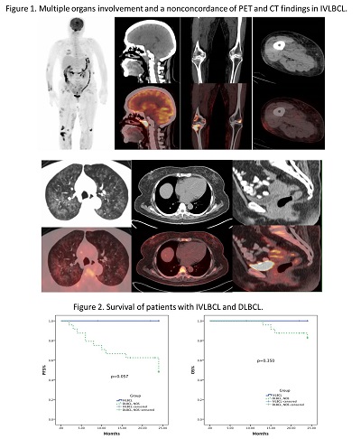
The characteristic PET/CT imaging of IVLBCL was summarized by comparing with DLBCL NOS. (Figure 1) IVLBCL involves multiple extranodal organs rather than lymph nodes, with a non concordance of PET and CT findings: 1). Patches without clear contour along the vessels traced by 18FDG; 2). Variable mild increase of SUVmax; 3). Normal CT imaging of solid organs; 4). Thickening subcutaneous/submucosal tissue or interstitial of the lung. All IVLBCL patients were diagnosed with the PET/CT directed biopsy, which led to a similar duration from the first symptom to diagnosis with those patients with DLBCL NOS (2.5 (1-10) vs. 2.5(1-12) months, P=0.798). No patients had concomitant hemophagocytic lymphohistiocytosis in both cohorts. Patients with IVLBCL achieved a complete metabolic response at both interim and at the end of treatment. With comparable IPI, COO and the time from diagnosis to treatment, there was a trend that patients with IVLBCL had superior progression-free survival than those with DLBCL stage IV (2-year PFS 100% vs. 50%, P= 0.057). (Figure 2)
Conclusion
A PET/CT-directed biopsy may be helpful for an early diagnosis of IVLBCL. Earlier chemoimmunotherapy may improve the prognosis of IVLBCL.
Keyword(s): B cell lymphoma, PET
Abstract: EP560
Type: E-Poster Presentation
Session title: Aggressive Non-Hodgkin lymphoma - Clinical
Background
Intravascular large B cell lymphoma (IVLBCL) is a rare and aggressive lymphoma. Early diagnosis of IVLBCL is difficult. Random skin biopsies and repetitive bone marrow biopsies are recommended to improve discrimination. Few studies focus on feasibility of PET/CT directed diagnosing in IVLBCL.
Aims
We conducted a single-center retrospective case-control study to explore the significance of PET/CT scans in patients with IVLBCL.
Methods
Data of consecutive 6 patients with IVLBCL from October 2018 to May 2020 were analyzed. 24 cases were extracted from patients with DLBCL NOS stage IV admitted and received treatment during the same period at a ratio of 4:1. The age of the patient in both IVLBCL and DLBCL cohorts was well matched. 5 patients in IVLBCL cohort received R-CHOP for 8 cycles and high-dose MTX (HD-MTX) for 2 cycles. 1 had the central nervous system (CNS) involvement besides systemic disorder at the diagnosis received autologous transplantation after 6 cycles of R-CHOP and 2 cycles of HD-MTX. 24 patients in DLBCL cohort received 8 cycles of R-CHOP. Patients who were at a high risk of secondary CNS lymphoma stratified with CNS-IPI or had involvement of sinus, bone marrow, adrenal glands or kidney received 2 cycles of HD-MTX. Patients were evaluated with PET/CT scan at diagnosis, interim and at the end of treatment. A comparison of clinical characteristics, PET/CT findings and outcome of patients in both cohorts was conducted.
Results
The characteristic PET/CT imaging of IVLBCL was summarized by comparing with DLBCL NOS. (Figure 1) IVLBCL involves multiple extranodal organs rather than lymph nodes, with a non concordance of PET and CT findings: 1). Patches without clear contour along the vessels traced by 18FDG; 2). Variable mild increase of SUVmax; 3). Normal CT imaging of solid organs; 4). Thickening subcutaneous/submucosal tissue or interstitial of the lung. All IVLBCL patients were diagnosed with the PET/CT directed biopsy, which led to a similar duration from the first symptom to diagnosis with those patients with DLBCL NOS (2.5 (1-10) vs. 2.5(1-12) months, P=0.798). No patients had concomitant hemophagocytic lymphohistiocytosis in both cohorts. Patients with IVLBCL achieved a complete metabolic response at both interim and at the end of treatment. With comparable IPI, COO and the time from diagnosis to treatment, there was a trend that patients with IVLBCL had superior progression-free survival than those with DLBCL stage IV (2-year PFS 100% vs. 50%, P= 0.057). (Figure 2)
Conclusion
A PET/CT-directed biopsy may be helpful for an early diagnosis of IVLBCL. Earlier chemoimmunotherapy may improve the prognosis of IVLBCL.
Keyword(s): B cell lymphoma, PET