

Contributions
Abstract: PB1770
Type: Publication Only
Session title: Sickle cell disease
Background
Sickle β-thalassemia (Sβ-thal) is a hereditary hemoglobinopathy resulting from the combined heterozygosity for sickle cell and β-thalassemia genes. Cardiac involvement in Sβ-thal patients has been poorly investigated.
Aims
We aimed to evaluate myocardial iron overload and cardiac function by cardiovascular magnetic resonance (CMR) in patients with Sβ-thal.
Methods
One hundred and eleven Sβ-thal patients consecutively enrolled in the Myocardial Iron Overload in Thalassemia (MIOT) network were studied and compared with 46 sickle cell disease (SCD) patients. Bi-atrial and biventricular function CMR parameters of Sβ-thal patients were compared with those of 111 healthy volunteers, matched by gender and age. Myocardial iron overload (MIO) was assessed by T2* technique. Cine images were acquired to quantify biventricular function. Macroscopic myocardial fibrosis was evaluated by late gadolinium enhancement (LGE) technique.
Results
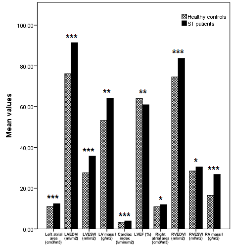
In Sβ-thal and SCD patients morphological and functional MR parameters were not significantly different, except for left atrial area and SVI (p = 0.023 and p = 0.048, respectively) that were significantly higher in SCD patients. No significant differences between the two groups were found in terms of myocardial iron overload and macroscopic myocardial fibrosis. When compared to healthy subjects, Sβ-thal patients showed significantly higher bi-atrial and biventricular parameters except for LVEF that was significantly lower (Fig.1).
Conclusion
The CMR analysis confirmed that Sβ-thal and SCD patients are phenotypically similar. Since Sβ-thal patients showed markedly different morphological and functional indices from healthy subjects, it would be useful to identify Sβ-thal/SCD-specific bi-atrial and biventricular reference values.
Keyword(s): Iron, Magnetic resonance imaging, Sickle cell anemia
Abstract: PB1770
Type: Publication Only
Session title: Sickle cell disease
Background
Sickle β-thalassemia (Sβ-thal) is a hereditary hemoglobinopathy resulting from the combined heterozygosity for sickle cell and β-thalassemia genes. Cardiac involvement in Sβ-thal patients has been poorly investigated.
Aims
We aimed to evaluate myocardial iron overload and cardiac function by cardiovascular magnetic resonance (CMR) in patients with Sβ-thal.
Methods
One hundred and eleven Sβ-thal patients consecutively enrolled in the Myocardial Iron Overload in Thalassemia (MIOT) network were studied and compared with 46 sickle cell disease (SCD) patients. Bi-atrial and biventricular function CMR parameters of Sβ-thal patients were compared with those of 111 healthy volunteers, matched by gender and age. Myocardial iron overload (MIO) was assessed by T2* technique. Cine images were acquired to quantify biventricular function. Macroscopic myocardial fibrosis was evaluated by late gadolinium enhancement (LGE) technique.
Results
In Sβ-thal and SCD patients morphological and functional MR parameters were not significantly different, except for left atrial area and SVI (p = 0.023 and p = 0.048, respectively) that were significantly higher in SCD patients. No significant differences between the two groups were found in terms of myocardial iron overload and macroscopic myocardial fibrosis. When compared to healthy subjects, Sβ-thal patients showed significantly higher bi-atrial and biventricular parameters except for LVEF that was significantly lower (Fig.1).
Conclusion
The CMR analysis confirmed that Sβ-thal and SCD patients are phenotypically similar. Since Sβ-thal patients showed markedly different morphological and functional indices from healthy subjects, it would be useful to identify Sβ-thal/SCD-specific bi-atrial and biventricular reference values.
Keyword(s): Iron, Magnetic resonance imaging, Sickle cell anemia