

Contributions
Abstract: PB1459
Type: Publication Only
Session title: Aggressive Non-Hodgkin lymphoma - Clinical
Background
Secondary central nervous system involvement as a relapse of primary mediastinal large B-cell lymphoma (PMLBCL) is an infrequent complication that drastically diminishes survival of the affected patients. Whole-brain radiotherapy and high-dose methotrexate (HD-MTX) were reported to improve the disease outcome, nevertheless, the median overall survival varies from 2 to 19 months, according to different authors. Here we present a case of complete cure of this condition.
Aims
The aim of the study was to outline the features of the disease course and management that could lead to long-term survival in case of primary mediastinal large B-cell lymphoma with secondary central nervous system involvement.
Methods
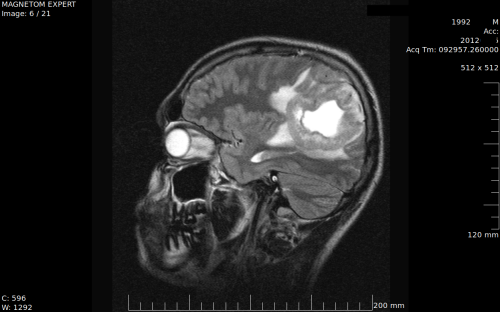
The 19-years-old male patient developed dyspnea and pain in the shoulder in June 2011. There were palpable subclavicular and left axillar lymph nodes revealed, and CT showed conglomerate in the mediastinum sized 160x135x190 mm together with contrast accumulation in both lungs and pleural effusion. Histological and immunohistochemical findings supported the diagnosis of PMLBCL. IPI was considered as low-intermediate. Initially patient received 2 CHOP-Bleo followed by 6 R-CHOP cycles, resulting in CR. Initially patient had received 2 CHOP-Bleo cycles of chemotherapy, followed by 6 R-CHOP cycles resulting in CR. One month later, however, intensive headaches developed. MRI revealed 71x62mm tumor in the right hemisphere (see picture). Right craniotomy with subtotal tumor resection had been performed, followed by 50 Gy 60Co γ-therapy that was used locally on the bed of the removed tumor with subsequent 42 Gy total brain irradiation. After resolution of cytopenia, 2 cycles of HD-MTX 3.5g/m2 and rituximab 0.375g/m2 were initiated with 3-week interval between cycles. Immunochemotherapy had been stopped due to the acquisition of acute viral hepatis B, and therapy with peg-interferon-α-2b (peg-IFN) 0.12g weekly was started and lasted 6 months.
Results
The decision to perform subtotal CNS tumor resection instead of biopsy was made intraoperatively by the surgeon, as the result of the macroscopical resemblance of the tumor to glioblastoma. Nevertheless, histology confirmed extranodal PMLBCL in the resected tissue. In the post-operative period, the patient developed secondary epilepsy, successfully controlled with carbamazepine. HD-MTX caused neutropenia with mild infectious complications. The switch from peg-IFN to tenofovir in the treatment of hepatitis B resulted in improvement of the symptoms of epilepsy. Residual dysmorphic changes in the right hemisphere have diminished spontaneously on the further MRI examinations. Body PET-CT after the stop of peg-IFN showed metabolically negative residual tumor mass in the mediastinum, sized 37x24x36mm. Biopsy revealed no bone marrow infiltration with PMLBCL. On the last PET-CT, performed 5 years after the CNS relapse, mediastinal residual tissue was slightly decreased to the maximal size of 29 mm. After the combined therapy the patient remains disease-free for 97 months. He has healthy biological son, born 3 years after the last cycle of HD-MTX.
Conclusion
Combination therapy including both surgical subtotal resection of localized CNS tumor instead of biopsy and subsequent local and whole brain irradiation followed by HD-MTX-containing regimens, can lead to the long-term remission and could possibly be curative for CNS relapse in some patients with PMLBCL. Further studies are needed to confirm the feasibility of this strategy.
Keyword(s): CNS lymphoma, High dose methotrexate, Radiotherapy, Surgery
Abstract: PB1459
Type: Publication Only
Session title: Aggressive Non-Hodgkin lymphoma - Clinical
Background
Secondary central nervous system involvement as a relapse of primary mediastinal large B-cell lymphoma (PMLBCL) is an infrequent complication that drastically diminishes survival of the affected patients. Whole-brain radiotherapy and high-dose methotrexate (HD-MTX) were reported to improve the disease outcome, nevertheless, the median overall survival varies from 2 to 19 months, according to different authors. Here we present a case of complete cure of this condition.
Aims
The aim of the study was to outline the features of the disease course and management that could lead to long-term survival in case of primary mediastinal large B-cell lymphoma with secondary central nervous system involvement.
Methods
The 19-years-old male patient developed dyspnea and pain in the shoulder in June 2011. There were palpable subclavicular and left axillar lymph nodes revealed, and CT showed conglomerate in the mediastinum sized 160x135x190 mm together with contrast accumulation in both lungs and pleural effusion. Histological and immunohistochemical findings supported the diagnosis of PMLBCL. IPI was considered as low-intermediate. Initially patient received 2 CHOP-Bleo followed by 6 R-CHOP cycles, resulting in CR. Initially patient had received 2 CHOP-Bleo cycles of chemotherapy, followed by 6 R-CHOP cycles resulting in CR. One month later, however, intensive headaches developed. MRI revealed 71x62mm tumor in the right hemisphere (see picture). Right craniotomy with subtotal tumor resection had been performed, followed by 50 Gy 60Co γ-therapy that was used locally on the bed of the removed tumor with subsequent 42 Gy total brain irradiation. After resolution of cytopenia, 2 cycles of HD-MTX 3.5g/m2 and rituximab 0.375g/m2 were initiated with 3-week interval between cycles. Immunochemotherapy had been stopped due to the acquisition of acute viral hepatis B, and therapy with peg-interferon-α-2b (peg-IFN) 0.12g weekly was started and lasted 6 months.
Results
The decision to perform subtotal CNS tumor resection instead of biopsy was made intraoperatively by the surgeon, as the result of the macroscopical resemblance of the tumor to glioblastoma. Nevertheless, histology confirmed extranodal PMLBCL in the resected tissue. In the post-operative period, the patient developed secondary epilepsy, successfully controlled with carbamazepine. HD-MTX caused neutropenia with mild infectious complications. The switch from peg-IFN to tenofovir in the treatment of hepatitis B resulted in improvement of the symptoms of epilepsy. Residual dysmorphic changes in the right hemisphere have diminished spontaneously on the further MRI examinations. Body PET-CT after the stop of peg-IFN showed metabolically negative residual tumor mass in the mediastinum, sized 37x24x36mm. Biopsy revealed no bone marrow infiltration with PMLBCL. On the last PET-CT, performed 5 years after the CNS relapse, mediastinal residual tissue was slightly decreased to the maximal size of 29 mm. After the combined therapy the patient remains disease-free for 97 months. He has healthy biological son, born 3 years after the last cycle of HD-MTX.
Conclusion
Combination therapy including both surgical subtotal resection of localized CNS tumor instead of biopsy and subsequent local and whole brain irradiation followed by HD-MTX-containing regimens, can lead to the long-term remission and could possibly be curative for CNS relapse in some patients with PMLBCL. Further studies are needed to confirm the feasibility of this strategy.
Keyword(s): CNS lymphoma, High dose methotrexate, Radiotherapy, Surgery