

Contributions
Abstract: PB2209
Type: Publication Only
Background
Cytogenetic study/ Fluorescence in situ Hybridization (FISH) has become an essential tool in assessing prognosis and survival in patients newly diagnosed with multiple myeloma. Cytogenetic abnormalities have various implications in terms of treatment response. It would be useful to identify patients with a higher incidence of high risk cytogenetic abnormalities in order to target further research in the vulnerable population.
Aims
To review distribution of cytogenetics abnormalities by FISH testing in different age groups of newly diagnosed multiple myeloma patients seen in the University Hospitals of Leicester NHS Trust between 2015-2018.
Methods
Data was collected from hospital computer-based patient records and the database of the cytogenetics department. All newly diagnosed patients with multiple myeloma who underwent a bone marrow biopsy were included in this analysis. . The bone marrow aspirates were screened morphologically. If there were ≥10% of plasma cells, the sample was referred for cytogenetics testing. The FISH panels consist of t (4;14), t (14;16), t (14;20), Del 17p, 1q amplification, and deletion 1p.
Results
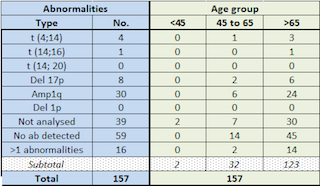
157 patients (92 male and 65 female) with newly diagnosed multiple myeloma underwent bone marrow biopsy over the three-year period. 39/157 were not analysed as either the plasma cells on the screening sample was insufficient or the number of cells cultured were inadequate for analysis. 59/157 did not have any FISH abnormality. 59/ 157 patients were tested positive for FISH abnormalities, out of which 16/59 had more than one FISH abnormality. The results were categorised under three age groups (< 45 years, 45-65 years and > 65 years). None of the patients under 45 had an abnormal FISH result while 11/32 patients between the ages of 45-65 and 48 /123 in the >65 year-old age group were identified with FISH abnormalities. . The most common FISH anomalies were amplification 1q followed by del 17P in both the intermediate age group and the older age population.
Conclusion
In our study it was observed that there was an increasing frequency of high risk mutations with increasing age . There is also a significantly higher proportion of del 1q seen in this UK cohort, this being a well known poor prognostic indicator. Cancer research UK data shows poorer patient survival ,shorter time to progression and poorer outcomes in this age group. More research needs to be targeted at the elderly population which may not be suited for more intensive therapies, to overcome high risk cytogenetics; but may benefit from targeted therapies given as maintenance treatment which may prevent progression and ensure better quality of life.
Session topic: 14. Myeloma and other monoclonal gammopathies - Clinical
Keyword(s): Cytogenetics, Multiple Myeloma
Abstract: PB2209
Type: Publication Only
Background
Cytogenetic study/ Fluorescence in situ Hybridization (FISH) has become an essential tool in assessing prognosis and survival in patients newly diagnosed with multiple myeloma. Cytogenetic abnormalities have various implications in terms of treatment response. It would be useful to identify patients with a higher incidence of high risk cytogenetic abnormalities in order to target further research in the vulnerable population.
Aims
To review distribution of cytogenetics abnormalities by FISH testing in different age groups of newly diagnosed multiple myeloma patients seen in the University Hospitals of Leicester NHS Trust between 2015-2018.
Methods
Data was collected from hospital computer-based patient records and the database of the cytogenetics department. All newly diagnosed patients with multiple myeloma who underwent a bone marrow biopsy were included in this analysis. . The bone marrow aspirates were screened morphologically. If there were ≥10% of plasma cells, the sample was referred for cytogenetics testing. The FISH panels consist of t (4;14), t (14;16), t (14;20), Del 17p, 1q amplification, and deletion 1p.
Results
157 patients (92 male and 65 female) with newly diagnosed multiple myeloma underwent bone marrow biopsy over the three-year period. 39/157 were not analysed as either the plasma cells on the screening sample was insufficient or the number of cells cultured were inadequate for analysis. 59/157 did not have any FISH abnormality. 59/ 157 patients were tested positive for FISH abnormalities, out of which 16/59 had more than one FISH abnormality. The results were categorised under three age groups (< 45 years, 45-65 years and > 65 years). None of the patients under 45 had an abnormal FISH result while 11/32 patients between the ages of 45-65 and 48 /123 in the >65 year-old age group were identified with FISH abnormalities. . The most common FISH anomalies were amplification 1q followed by del 17P in both the intermediate age group and the older age population.
Conclusion
In our study it was observed that there was an increasing frequency of high risk mutations with increasing age . There is also a significantly higher proportion of del 1q seen in this UK cohort, this being a well known poor prognostic indicator. Cancer research UK data shows poorer patient survival ,shorter time to progression and poorer outcomes in this age group. More research needs to be targeted at the elderly population which may not be suited for more intensive therapies, to overcome high risk cytogenetics; but may benefit from targeted therapies given as maintenance treatment which may prevent progression and ensure better quality of life.
Session topic: 14. Myeloma and other monoclonal gammopathies - Clinical
Keyword(s): Cytogenetics, Multiple Myeloma