

Contributions
Abstract: PB2119
Type: Publication Only
Background
Multiple myeloma (MM) is the second most prevalent hematologic malignancy after non-Hodgkin’s lymphoma. Drug resistance and relapse limit MM to be an incurable disease. Mogrosides are reported to represent a novel class of bioactive plant compounds with anti-cancer effects, which are digested to mogrol in vivo.
Aims
Here, we sought to verify the anti-myeloma effect of mogrol both in vitro and in vivo, and investigate the pharmacological mechanism to provide new therapeutic strategy for MM.
Methods
Inhibitory rates under different concentrations of mogrol between 0 and 50umol/L were measured via cell count kit-8 (CCK-8) to ascertain the maximal inhibitory concentration (IC50). Peripheral blood mononuclear cells (PBMCs) from health donors was used to make sure the safety of mogrol. Next, 2 myeloma cell lines (ARH77 and U266) were exposed to mogrol at different concentrations of 0, 0.5×IC50 and IC50, and cell viability, apoptosis and cell cycle distribution were measured to verify the anti-cancer effects of mogrol on MM cells. Moreover, myeloma xenografted BALB/c nude mice model was established to validate the pharmacological effects of mogrol in vivo. The Raf/MEK/extracellular regulated kinase (ERK) and nuclear factor kappa-light-chain-enhancer of activated B cells (NF-κB) pathways were investigated through detecting related proteins expression by western blot.
Results
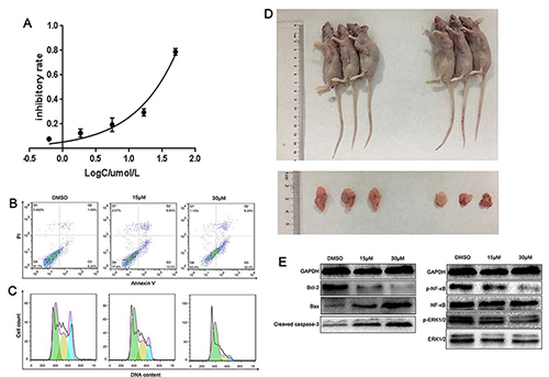
CCK-8 test showed that mogrol significant inhibited MM cell proliferation in a time- and dose-dependent manner. However, viability of PBMCs was not influenced by mogrol at any concentrations. And the inhibitory rate on MM cells changed from 7.73% to 78.9% at concentrations between 0.62 and 50umol/L, thus IC50 of mogrol on myeloma cells was 26.52±0.21umol/L. In addition, we also found that mogrol not only decreased MM cell viability, increased the total apoptosis rate, and caused G1/S arrest in vitro, but also reduced tumor volume and slowed down tumor growth in vivo, with an average size of tumors of 228.20±11.02mm3 vs. 422.90±50.07mm3 after 4 weeks (p< 0.05). Accordingly, western blot results displayed the dysregulation of relative proteins expression, such as up-regulated Bax and Cleaved caspase-3, and down-regulated Bcl-2. Moreover, ERK phosphorylation was inhibited in MM cells treated with mogrol, and NF-κB was also decreased in the mogrol-treated MM cells.
Legends:
A. Inhibitory effect of mogrol on ARH-77 cell proliferation after 24h.
B. Apoptotic effects of mogrol on ARH-77 cells as determined by flow cytometry.
C. Mogrol induced G1/S phase arrest in ARH-77 cells.
D. Mogrol inhibited tumor growth in the myeloma xenografted BALB/c nude mice model.
E. Effects of mogrol on the expression of p-NF-κB, p-ERK1/2, Bcl-2, Bax, and Cleaved caspase-3.
Conclusion
Our results showed that mogrol not only inhibited MM cells growth in vitro, but also hampered MM development and progression in vivo. Besides, we found that ERK and NF-κB pathways was inhibited in the MM cells treated with mogrol. These findings suggested that mogrol, a novel ERK inhibiting agents, might grow to be a safe and promising anti-myeloma drug.
Session topic: 13. Myeloma and other monoclonal gammopathies – Biology & Translational Research
Keyword(s): Drug interaction, ERK, Multiple Myeloma, NF- B
Abstract: PB2119
Type: Publication Only
Background
Multiple myeloma (MM) is the second most prevalent hematologic malignancy after non-Hodgkin’s lymphoma. Drug resistance and relapse limit MM to be an incurable disease. Mogrosides are reported to represent a novel class of bioactive plant compounds with anti-cancer effects, which are digested to mogrol in vivo.
Aims
Here, we sought to verify the anti-myeloma effect of mogrol both in vitro and in vivo, and investigate the pharmacological mechanism to provide new therapeutic strategy for MM.
Methods
Inhibitory rates under different concentrations of mogrol between 0 and 50umol/L were measured via cell count kit-8 (CCK-8) to ascertain the maximal inhibitory concentration (IC50). Peripheral blood mononuclear cells (PBMCs) from health donors was used to make sure the safety of mogrol. Next, 2 myeloma cell lines (ARH77 and U266) were exposed to mogrol at different concentrations of 0, 0.5×IC50 and IC50, and cell viability, apoptosis and cell cycle distribution were measured to verify the anti-cancer effects of mogrol on MM cells. Moreover, myeloma xenografted BALB/c nude mice model was established to validate the pharmacological effects of mogrol in vivo. The Raf/MEK/extracellular regulated kinase (ERK) and nuclear factor kappa-light-chain-enhancer of activated B cells (NF-κB) pathways were investigated through detecting related proteins expression by western blot.
Results
CCK-8 test showed that mogrol significant inhibited MM cell proliferation in a time- and dose-dependent manner. However, viability of PBMCs was not influenced by mogrol at any concentrations. And the inhibitory rate on MM cells changed from 7.73% to 78.9% at concentrations between 0.62 and 50umol/L, thus IC50 of mogrol on myeloma cells was 26.52±0.21umol/L. In addition, we also found that mogrol not only decreased MM cell viability, increased the total apoptosis rate, and caused G1/S arrest in vitro, but also reduced tumor volume and slowed down tumor growth in vivo, with an average size of tumors of 228.20±11.02mm3 vs. 422.90±50.07mm3 after 4 weeks (p< 0.05). Accordingly, western blot results displayed the dysregulation of relative proteins expression, such as up-regulated Bax and Cleaved caspase-3, and down-regulated Bcl-2. Moreover, ERK phosphorylation was inhibited in MM cells treated with mogrol, and NF-κB was also decreased in the mogrol-treated MM cells.
Legends:
A. Inhibitory effect of mogrol on ARH-77 cell proliferation after 24h.
B. Apoptotic effects of mogrol on ARH-77 cells as determined by flow cytometry.
C. Mogrol induced G1/S phase arrest in ARH-77 cells.
D. Mogrol inhibited tumor growth in the myeloma xenografted BALB/c nude mice model.
E. Effects of mogrol on the expression of p-NF-κB, p-ERK1/2, Bcl-2, Bax, and Cleaved caspase-3.
Conclusion
Our results showed that mogrol not only inhibited MM cells growth in vitro, but also hampered MM development and progression in vivo. Besides, we found that ERK and NF-κB pathways was inhibited in the MM cells treated with mogrol. These findings suggested that mogrol, a novel ERK inhibiting agents, might grow to be a safe and promising anti-myeloma drug.
Session topic: 13. Myeloma and other monoclonal gammopathies – Biology & Translational Research
Keyword(s): Drug interaction, ERK, Multiple Myeloma, NF- B