

Contributions
Abstract: PB2142
Type: Publication Only
Background
Multiple myeloma is a B cell neoplastic disorder characterized by clonal proliferation of malignant plasma cells in the bone marrow. CD138 is a marker of plasma cells and MM cells. The expression of CD138 highly correlates with that of VEGFR3. However, a low CD138 expression is frequently observed in MM patients and several studies have reported how a decreased CD138 expression can affect the prognosis and the treatment response.
Aims
We proposed Micro-Raman spectroscopy as an easily accurate and non invasive method to discriminate plasma cell immune-phenotype.
Methods
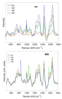
two distinct cell subtypes for antigen expression pattern: i) subtype 2 CD45-/CD56-/CD38+ (namely, S01); ii) subtype 3 CD45+/CD38+/CD138+ (namely, S02) separated by the conventional flow cytometry approach and then fixed on CaF2 substrate, were analyzed carrying out Raman mapping analyses. Raman contributions were observed in selected spectral regions where markers of specific functional groups, useful to characterize the cell state, are present. Hence, comparing Raman spectra, the investigated cell subtypes can be distinguished, analyzing several spectra for each sample. So, intracellular variability, repeatability and sensitivity of the analyses for each sample was evaluated.
Results
- Regarding S01cells: the spectra acquired in the #3 and #4 points are characterized by DNA and PO-P vibrational modes in the 637-665 and 776-827 cm-1, respectively. In addition, RNA features at 725 cm-1 and in the 1560-1580 cm-1 regions are more evident. C-C or C-O vibrational modes, ascribed to membrane phospholipids, are envisaged in the 1075-1116 cm-1 region.
- Regarding S02 cells: The main spectral differences respect to S01 cells are referred: 1) to the nucleic acids contributions (DNA/RNA) and 2) to the protein features (1255-1670 cm-1 region) more pronounced and defined in the S02 cells in comparison to the S01 ones. Furthermore, cells S01 show an high internal variability respect to S02, suggesting a high metabolic activity.
Conclusion
Plasma cells are a eterogenous population in Multiple Myeloma patients. A different rate of CD138+ cells is able to modify the prognosis. Raman Spectroscopy could increase our knowledge of the structure and problably the function of these cells contribuiting to a better awereness of the mechanisms of Multiple Myeloma progression.
Session topic: 13. Myeloma and other monoclonal gammopathies – Biology & Translational Research
Keyword(s): Multiple Myeloma, Plasma cells, prognosis
Abstract: PB2142
Type: Publication Only
Background
Multiple myeloma is a B cell neoplastic disorder characterized by clonal proliferation of malignant plasma cells in the bone marrow. CD138 is a marker of plasma cells and MM cells. The expression of CD138 highly correlates with that of VEGFR3. However, a low CD138 expression is frequently observed in MM patients and several studies have reported how a decreased CD138 expression can affect the prognosis and the treatment response.
Aims
We proposed Micro-Raman spectroscopy as an easily accurate and non invasive method to discriminate plasma cell immune-phenotype.
Methods
two distinct cell subtypes for antigen expression pattern: i) subtype 2 CD45-/CD56-/CD38+ (namely, S01); ii) subtype 3 CD45+/CD38+/CD138+ (namely, S02) separated by the conventional flow cytometry approach and then fixed on CaF2 substrate, were analyzed carrying out Raman mapping analyses. Raman contributions were observed in selected spectral regions where markers of specific functional groups, useful to characterize the cell state, are present. Hence, comparing Raman spectra, the investigated cell subtypes can be distinguished, analyzing several spectra for each sample. So, intracellular variability, repeatability and sensitivity of the analyses for each sample was evaluated.
Results
- Regarding S01cells: the spectra acquired in the #3 and #4 points are characterized by DNA and PO-P vibrational modes in the 637-665 and 776-827 cm-1, respectively. In addition, RNA features at 725 cm-1 and in the 1560-1580 cm-1 regions are more evident. C-C or C-O vibrational modes, ascribed to membrane phospholipids, are envisaged in the 1075-1116 cm-1 region.
- Regarding S02 cells: The main spectral differences respect to S01 cells are referred: 1) to the nucleic acids contributions (DNA/RNA) and 2) to the protein features (1255-1670 cm-1 region) more pronounced and defined in the S02 cells in comparison to the S01 ones. Furthermore, cells S01 show an high internal variability respect to S02, suggesting a high metabolic activity.
Conclusion
Plasma cells are a eterogenous population in Multiple Myeloma patients. A different rate of CD138+ cells is able to modify the prognosis. Raman Spectroscopy could increase our knowledge of the structure and problably the function of these cells contribuiting to a better awereness of the mechanisms of Multiple Myeloma progression.
Session topic: 13. Myeloma and other monoclonal gammopathies – Biology & Translational Research
Keyword(s): Multiple Myeloma, Plasma cells, prognosis