

Contributions
Abstract: PB2133
Type: Publication Only
Background
At the time of the diagnosis of multiple myeloma (MM) the incidence of plasmacytomas varies from 3,5 to 18%. We defined plasmacytomas as bone (plasma cell tumor extending from the intramedullary spaces of the bone marrow through the cortical bone) and extramedullary (spread to organs distant from bone). The presence of extramedullary plasmacytoma at the time of the diagnosis of MM has a negative impact on the disease prognosis, while bone plasmacytomas in patients with MM are treatable. The pathogenesis of extramedullary disease has not been thoroughly analyzed, thought cell adhesion molecules and chemokine receptors are assumed to participate in this process. Study of tumor plasmacytoma substrate is great interest due to the rarity of this pathology.
Aims
Analyzing CD56, CD166, CXCR4, Ki-67 and c-MYC expression in the tumor substrate of bone and extramedullary plasmacytomas in patients with MM.
Methods
From October 2013 to April 2017 21 patients with newly diagnosed MM (10 males and 11 females) 23 - 77 years old (mean value: 52 y.o.) were included in the study. The disease was diagnosed in accordance to IMWG criteria (2014). 14 patients were diagnosed with bone plasmacytoma associated with skeleton bones (vertebrae, ribs, skull and pelvis) and 7 patients were diagnosed with extramedullary plasmacytoma distant from bone (liver, pancreas, stomach, soft tissues, muscles and skin). In all cases a tumour biopsy was taken which confirmed the presence of plasma cell infiltration. Paraffin block slices from tumour biopsy material were used to perform an immunohistochemistry analysis with an antibody panel to CD56, Ki67, CXCR4, CD166, c-MYC. Marker expression level was analyzed with Leica DM4000B microscope by viewing 10 fields of view at 400-fold magnification. Marker expression assessment was carried out by means of semiquantitative method. The percentage of cells expressing the protein in question against the total number of tumor substrate cells was calculated. To assess statistical differences between the mean values we applied Student’s t-test (including preliminary assessment using Levene’s test for equality of variances).
Results
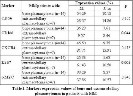
Analyzing mean values of marker expression in bone and extramedullary plasmacytomas resulted in revealing significant differences in CD166 and Кi-67 (Table 1), while no significant differences between expressions of other markers in question were found. A higher Ki-67 index (p=0.004) was reported for extramedullary plasmacytoma cells, comparing to that of bone plasmacytoma cells, with their mean values of 61.43±10.50% and 25.36±5.63% respectively. The expression of cell adhesion molecule CD166 in bone plasmacytoma cells was significantly higher than that of extramedullary plasmacytoma cells (36.29±7.61% and 9.57±8.46% respectively, p=0.044).
Conclusion
Aggressive course of MM complicated with extramedullary plasmacytoma may be caused by significantly higher proliferative activity of extramedullary plasmacytoma cells, comparing to that of bone plasmacytoma cells.
Low expression of adhesion molecule CD166 in extramedullary plasmacytoma cells may be the reason behind weakening bond between myeloma cell and bone marrow microenvironment, which leads to the occurrence of hematogenic dissemination of plasma cells in various organs and tissues. In the same time, high expression of CD166 in bone plasmacytoma cells may point to this marker’s participation in modelling osteogenesis and forming osteodestructions.
Session topic: 13. Myeloma and other monoclonal gammopathies – Biology & Translational Research
Keyword(s): Ki-67, Multiple Myeloma, Plasma cells
Abstract: PB2133
Type: Publication Only
Background
At the time of the diagnosis of multiple myeloma (MM) the incidence of plasmacytomas varies from 3,5 to 18%. We defined plasmacytomas as bone (plasma cell tumor extending from the intramedullary spaces of the bone marrow through the cortical bone) and extramedullary (spread to organs distant from bone). The presence of extramedullary plasmacytoma at the time of the diagnosis of MM has a negative impact on the disease prognosis, while bone plasmacytomas in patients with MM are treatable. The pathogenesis of extramedullary disease has not been thoroughly analyzed, thought cell adhesion molecules and chemokine receptors are assumed to participate in this process. Study of tumor plasmacytoma substrate is great interest due to the rarity of this pathology.
Aims
Analyzing CD56, CD166, CXCR4, Ki-67 and c-MYC expression in the tumor substrate of bone and extramedullary plasmacytomas in patients with MM.
Methods
From October 2013 to April 2017 21 patients with newly diagnosed MM (10 males and 11 females) 23 - 77 years old (mean value: 52 y.o.) were included in the study. The disease was diagnosed in accordance to IMWG criteria (2014). 14 patients were diagnosed with bone plasmacytoma associated with skeleton bones (vertebrae, ribs, skull and pelvis) and 7 patients were diagnosed with extramedullary plasmacytoma distant from bone (liver, pancreas, stomach, soft tissues, muscles and skin). In all cases a tumour biopsy was taken which confirmed the presence of plasma cell infiltration. Paraffin block slices from tumour biopsy material were used to perform an immunohistochemistry analysis with an antibody panel to CD56, Ki67, CXCR4, CD166, c-MYC. Marker expression level was analyzed with Leica DM4000B microscope by viewing 10 fields of view at 400-fold magnification. Marker expression assessment was carried out by means of semiquantitative method. The percentage of cells expressing the protein in question against the total number of tumor substrate cells was calculated. To assess statistical differences between the mean values we applied Student’s t-test (including preliminary assessment using Levene’s test for equality of variances).
Results
Analyzing mean values of marker expression in bone and extramedullary plasmacytomas resulted in revealing significant differences in CD166 and Кi-67 (Table 1), while no significant differences between expressions of other markers in question were found. A higher Ki-67 index (p=0.004) was reported for extramedullary plasmacytoma cells, comparing to that of bone plasmacytoma cells, with their mean values of 61.43±10.50% and 25.36±5.63% respectively. The expression of cell adhesion molecule CD166 in bone plasmacytoma cells was significantly higher than that of extramedullary plasmacytoma cells (36.29±7.61% and 9.57±8.46% respectively, p=0.044).
Conclusion
Aggressive course of MM complicated with extramedullary plasmacytoma may be caused by significantly higher proliferative activity of extramedullary plasmacytoma cells, comparing to that of bone plasmacytoma cells.
Low expression of adhesion molecule CD166 in extramedullary plasmacytoma cells may be the reason behind weakening bond between myeloma cell and bone marrow microenvironment, which leads to the occurrence of hematogenic dissemination of plasma cells in various organs and tissues. In the same time, high expression of CD166 in bone plasmacytoma cells may point to this marker’s participation in modelling osteogenesis and forming osteodestructions.
Session topic: 13. Myeloma and other monoclonal gammopathies – Biology & Translational Research
Keyword(s): Ki-67, Multiple Myeloma, Plasma cells