

Contributions
Abstract: PB1660
Type: Publication Only
Background
Acute leukemia diagnosis requires the integration of morphology and flow cytometry data. However, it is not always straightforward due to the difference in the method for blast identification used in both cases.
Aims
To simplify the integration of morphology and immunophenotyping we have developed an anti-cluster-of-differentiation (anti-CD) antibody microarray on a transparent support for leukocyte sorting and a method for preparation of the microarray-bound cells for high-resolution morphology analysis.
Methods
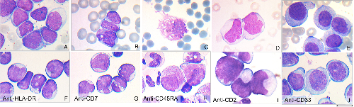
The suspension of mononuclear cells separated by density gradient from bone marrow aspirate is incubated with the microarray in non-mixing conditions. After the unbound cells are washed away the bound cells are dried and flattened in a home-developed cytocentrifuge. The drying/flattening procedure makes the microarray-bound cells morphologically identical to the same cells in a smear and suitable for other standard smear-oriented techniques such as cytochemistry while the leukocytes on the microarray are grouped according to their surface CD antigens. Due to the non-mixing incubation the density of the cells bound to an anti-CD antibody permits to determine the fraction of cells positive for the corresponding CD antigen with high correlation with flow cytometry.
Results
We studied 66 patients with diagnosed acute leukemia of different subtypes and showed that the morphology of the microarray-bound blast cells was identical to the same cells in smears, the percentage of blast cells was in good correlation with the results obtained by flow cytometry and smear analysis and the blast immunophenotype determined as the list of anti-CD antibodies binding the leukocyte population with more than 2% of cells with blast morphology agreed well with flow cytometry results. Then we have compared the blast percentage among anti-CD45-bound bone marrow mononuclear cells for 23 nonneoplastic bone marrow aspirates, 17 samples from patients with chronic myelocytic or myelomonocytic leukemia and 90 bone marrow aspirates from patients with acute leukemias. The threshold for acute leukemia was found to be 25%. Finally we show that morphology and cytochemistry analysis of mononuclear bone marrow cells from patients with suspected acute leukemia bound by anti-CD antibody microarray including monoclonal antibodies against CD1a, CD2, CD3, CD4, CD5, CD7, CD8, CD10, CD11b, CD11c, CD13, CD14, CD15, CD16, CD19, CD20, CD22, CD33, CD41, CD45, CD61, CD64, CD117, CD235, IgM, HLA-DR and negative control permits to arrive at preliminary diagnosis by FAB classification.
Conclusion
The anti-CD morphology microarray can help in integration of morphology and flow cytometry data in differential diagnosis of acute leukemias and is a potentially useful diagnostic tool in resource-poor countries.
The work was partially supported by gran #18-015-00272 from Russian Foundation for Basic Research.
Session topic: 3. Acute myeloid leukemia - Biology & Translational Research
Keyword(s): Acute Myeloid Leukemia, Diagnosis
Abstract: PB1660
Type: Publication Only
Background
Acute leukemia diagnosis requires the integration of morphology and flow cytometry data. However, it is not always straightforward due to the difference in the method for blast identification used in both cases.
Aims
To simplify the integration of morphology and immunophenotyping we have developed an anti-cluster-of-differentiation (anti-CD) antibody microarray on a transparent support for leukocyte sorting and a method for preparation of the microarray-bound cells for high-resolution morphology analysis.
Methods
The suspension of mononuclear cells separated by density gradient from bone marrow aspirate is incubated with the microarray in non-mixing conditions. After the unbound cells are washed away the bound cells are dried and flattened in a home-developed cytocentrifuge. The drying/flattening procedure makes the microarray-bound cells morphologically identical to the same cells in a smear and suitable for other standard smear-oriented techniques such as cytochemistry while the leukocytes on the microarray are grouped according to their surface CD antigens. Due to the non-mixing incubation the density of the cells bound to an anti-CD antibody permits to determine the fraction of cells positive for the corresponding CD antigen with high correlation with flow cytometry.
Results
We studied 66 patients with diagnosed acute leukemia of different subtypes and showed that the morphology of the microarray-bound blast cells was identical to the same cells in smears, the percentage of blast cells was in good correlation with the results obtained by flow cytometry and smear analysis and the blast immunophenotype determined as the list of anti-CD antibodies binding the leukocyte population with more than 2% of cells with blast morphology agreed well with flow cytometry results. Then we have compared the blast percentage among anti-CD45-bound bone marrow mononuclear cells for 23 nonneoplastic bone marrow aspirates, 17 samples from patients with chronic myelocytic or myelomonocytic leukemia and 90 bone marrow aspirates from patients with acute leukemias. The threshold for acute leukemia was found to be 25%. Finally we show that morphology and cytochemistry analysis of mononuclear bone marrow cells from patients with suspected acute leukemia bound by anti-CD antibody microarray including monoclonal antibodies against CD1a, CD2, CD3, CD4, CD5, CD7, CD8, CD10, CD11b, CD11c, CD13, CD14, CD15, CD16, CD19, CD20, CD22, CD33, CD41, CD45, CD61, CD64, CD117, CD235, IgM, HLA-DR and negative control permits to arrive at preliminary diagnosis by FAB classification.
Conclusion
The anti-CD morphology microarray can help in integration of morphology and flow cytometry data in differential diagnosis of acute leukemias and is a potentially useful diagnostic tool in resource-poor countries.
The work was partially supported by gran #18-015-00272 from Russian Foundation for Basic Research.
Session topic: 3. Acute myeloid leukemia - Biology & Translational Research
Keyword(s): Acute Myeloid Leukemia, Diagnosis