

Contributions
Abstract: PB2153
Type: Publication Only
Background
No data are available in literature about the relationship between gender and the development of CMR abnormalities and/or cardiac complications in sickle cell disease (SCD).
Aims
This prospective and multicentre study aimed to assess if there was an association between gender and risk of cardiac iron overload, heart dysfunction and dilation, left ventricular (LV) hypertrophy, and myocardial fibrosis, assessed by Cardiovascular Magnetic Resonance (CMR), and of cardio-vascular complications in sickle cell disease (SCD) patients.
Methods
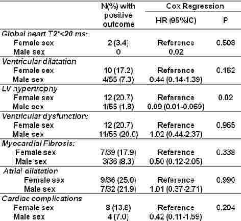
We considered 115 SCD patients (58 females, 34.79±13.26 years), consecutively enrolled in the Myocardial Iron Overload in Thalassemia (MIOT) Network. Myocardial iron overload was assessed by the multislice multiecho T2* technique. Biventricular function parameters and atrial areas were quantified by cine images. Late gadolinium enhancement (LGE) images were acquired to detect myocardial fibrosis.
Results
Conclusion
Session topic: 25. Sickle cell disease
Keyword(s): Magnetic resonance imaging, Gender
Abstract: PB2153
Type: Publication Only
Background
No data are available in literature about the relationship between gender and the development of CMR abnormalities and/or cardiac complications in sickle cell disease (SCD).
Aims
This prospective and multicentre study aimed to assess if there was an association between gender and risk of cardiac iron overload, heart dysfunction and dilation, left ventricular (LV) hypertrophy, and myocardial fibrosis, assessed by Cardiovascular Magnetic Resonance (CMR), and of cardio-vascular complications in sickle cell disease (SCD) patients.
Methods
We considered 115 SCD patients (58 females, 34.79±13.26 years), consecutively enrolled in the Myocardial Iron Overload in Thalassemia (MIOT) Network. Myocardial iron overload was assessed by the multislice multiecho T2* technique. Biventricular function parameters and atrial areas were quantified by cine images. Late gadolinium enhancement (LGE) images were acquired to detect myocardial fibrosis.
Results
Conclusion
Session topic: 25. Sickle cell disease
Keyword(s): Magnetic resonance imaging, Gender