EHA Library - The official digital education library of European Hematology Association (EHA)

FLUORESCENCE IN SITU HYBRIDIZATION SIGNAL PATTERNS AND INTRACHROMOSOMAL BCR-ABL1 AMPLIFICATION ANALYSIS IN IMATINIB-RESISTANT CHRONIC MYELOGENOUS LEUKEMIA PATIENTS USING TRICOLOR DUAL FUSION PROBE.
Author(s): ,
Karthik Bommannan
Affiliations:
Hematology,Post Graduate Institute of Medical Education and Research,CHANDIGARH,India;Hematology,Post Graduate Institute of Medical Education and Research,CHANDIGARH,India
,
SHANO NASEEM
Affiliations:
Hematology,Post Graduate Institute of Medical Education and Research,CHANDIGARH,India
,
NEELAM VARMA
Affiliations:
Hematology,Post Graduate Institute of Medical Education and Research,CHANDIGARH,India
,
PANKAJ MALHOTRA
Affiliations:
Internal Medicine,Post Graduate Institute of Medical Education and Research,CHANDIGARH,India
,
JOGESHWAR BINOTA
Affiliations:
Hematology,Post Graduate Institute of Medical Education and Research,CHANDIGARH,India
SUBHASH VARMA
Affiliations:
Internal Medicine,Post Graduate Institute of Medical Education and Research,CHANDIGARH,India
(Abstract release date: 05/18/17) EHA Library. Bommannan K. 05/18/17; 182519; PB1805
Karthik Bommannan
Karthik Bommannan
Contributions
Abstract

Abstract: PB1805

Type: Publication Only

Background
Conventional cytogenetics is a common modality for tyrosine kinase inhibitor (TKI) response assessment in chronic myelogenous leukemia (CML) patients. There is no consensus regarding the use of conventional bone marrow (BM) cytogenetics or peripheral blood (PB) interphase fluorescence in situ hybridization (I-FISH) during follow-up. The routine dual colour FISH probes are less sensitive to reliably identify der(9) deletions during follow-up. BCR/ABL/ASS1 tri-colour dual fusion (TCDF) probe is highly sensitive and specific in identifying der(9) deletions.

Aims
Our aim was to identify the I-FISH fusion patterns of BCR/ABL/ASS1 TCDF probe and correlate the patterns with patient-specific molecular genetic parameters.

Methods

This was an ethically approved study conducted at a government-funded tertiary care institute. From January 2015 to June 2016, PB I-FISH analysis was performed on European LeukemiaNet defined imatinib-resistant CML patients using BCR/ABL/ASS1 TCDF probe (Abbott Laboratories, Abbott Park, Illinois, USA). The residual BCR-ABL1 transcript load was monitored in international scale (BCR-ABL1IS) using an automated cartridge-based GeneXpert system (Cepheid, Sunnyvale, CA, USA).

Results

On analyzing 37 adult patients, all had residual Philadelphia (Ph) chromosomes (100%). Classic Ph fusion pattern was seen in 33 (89%), derivative chromosome 9 [der(9)] deletions in 25 (67.5%) and supernumerary Ph chromosomes in 11 (30%) patients. Coexistence of classical fusion and der(9) deletions were seen in 21 patients (57%), whereas 8 patients (22%) had a mutual existence of classical fusion, der(9) deletions and supernumerary Ph chromosomes. None had Ph amplification.
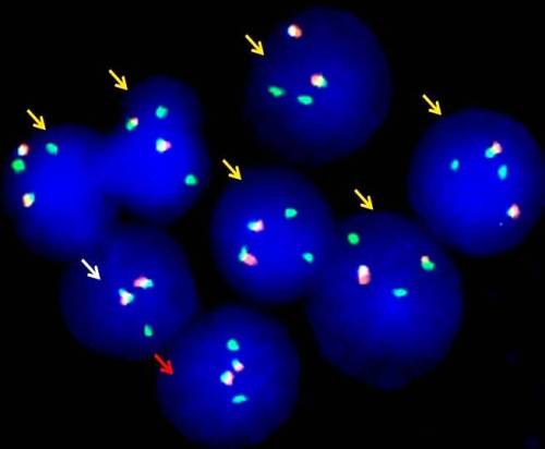
Figure 1 demonstrates the I-FISH patterns seen in a 43-year-old male diagnosed with CML-CP and had progressed to blast crisis at his 72nd month of imatinib therapy. In this figure red, yellow and white arrows indicate blast cells without Ph chromosome, Ph+ blast with a loss of residual ABL1 on der(9)classical and random signal overlap, respectively.
A mean (± S.D) of 29% (± 30) and 18% (± 17) der(9) deleted cells were seen amongst patients with b2a2 and b3a2 BCR-ABL1 transcript types, respectively and this difference was statistically significant (p=0.008). There was also a significant difference in the disease transformation status according to the percentage of der(9) deleted cells (p=0.03). In this regard, patients with progressive disease (accelerated phase/ blast crisis progression) had a mean (± S.D) of 47 % (± 35) der(9) deleted cells in comparison to 19% (± 20) such cells in patients without disease transformation. In addition, patients with Ph duplication/triplication had a mean (± S.D) BCR-ABL1IS levels of 49.478% (± 40.184), in comparison to BCR-ABL1IS levels of 16.00% (± 19.993) in patients without these anomalies and this difference was also statistically significant (p=0.029).

Conclusion
Our work would be an appropriate reference material for I-FISH signal interpretation using BCR/ABL/ASS1 TCDF probe. We have demonstrated a high frequency of der(9) deletions, clonal heterogeneity and absence of BCR-ABL1 amplification in an imatinib-resistant Indian CML cohort. For the first time, a significant association of der(9) deleted cell percentage with b2a2 transcript type and disease transformation status has been identified and the same has to be tested in a larger cohort.

Session topic: 7. Chronic myeloid leukemia - Biology

Keyword(s): Chronic myeloid leukemia, FISH

By clicking “Accept Terms & all Cookies” or by continuing to browse, you agree to the storing of third-party cookies on your device to enhance your user experience and agree to the user terms and conditions of this learning management system (LMS).

Cookie Settings
Accept Terms & all Cookies