

Contributions
Abstract: O06
Type: Oral presentation
Session Topic: Inherited and acquired coagulation disorders
Presentation during EHA Scientific Conference on Bleeding Disorders:
On Thursday, September 15, 2016 from 09:00 - 10:30
Location: Rubí + Zafir
Background
The calibrated automated thrombogram (CAT) assay is emerging as a reliable tool for real time estimation of thrombin generation (TG) potential. There is limited knowledge about the differences in the pathways that underlie the thrombotic phenotype in the solid versus haematological malignant condition. Few studies have investigated the contribution to thrombosis of different factors of the coagulation cascade1. Characterizing the TG capacity in these two distinct cancer models using factor deficient plasma might potentially allow better characterization of the thrombotic risk and individualization of prevention strategies.
Methods
Solid tumour cells (pancreatic cancer AsPC-1, CFPAC-1, PANC-1, MIA PaCa-2 and others such as SKOV-3 ovarian cancer, UMSCC81B Head & Neck squamous cancer, PC9 lung cancer) and malignant haematological cell lines (Multiple myeloma MM1.S, U266B, H929 and others including JJN3 plasma cell leukaemia, U937 histiocytic lymphoma) were evaluated at increasing cell concentrations on a Thrombinoscope for the CAT assay, with the addition of platelet-free normal plasma (NormTrol) or plasma deficient in coagulation factors VII and XII, and TF 1pM standard preparations as control. In addition, tissue factor (TF) cell surface expression was measured with flow cytometry.
Results
In NormTrol plasma, TG in all cancer cell lines was concentration dependent, with CFPAC-1 producing the highest thrombin. Absence of Factor-VII in platelet-free plasma resulted in significantly higher inhibition of TG in solid cancers compared to haematological cancers (Figure 2). TF surface expression correlated strongly with the TG parameters e.g. the higher the TF expressed in a cell line, then the shorter the Lag time and time-to-peak, and UMSCC81B expressed the highest TF. Compared to 1pM TF control, solid tumour cell lines had higher thrombin peaks, faster lag times, and a TG profile of overall greater magnitude than haematological cell lines.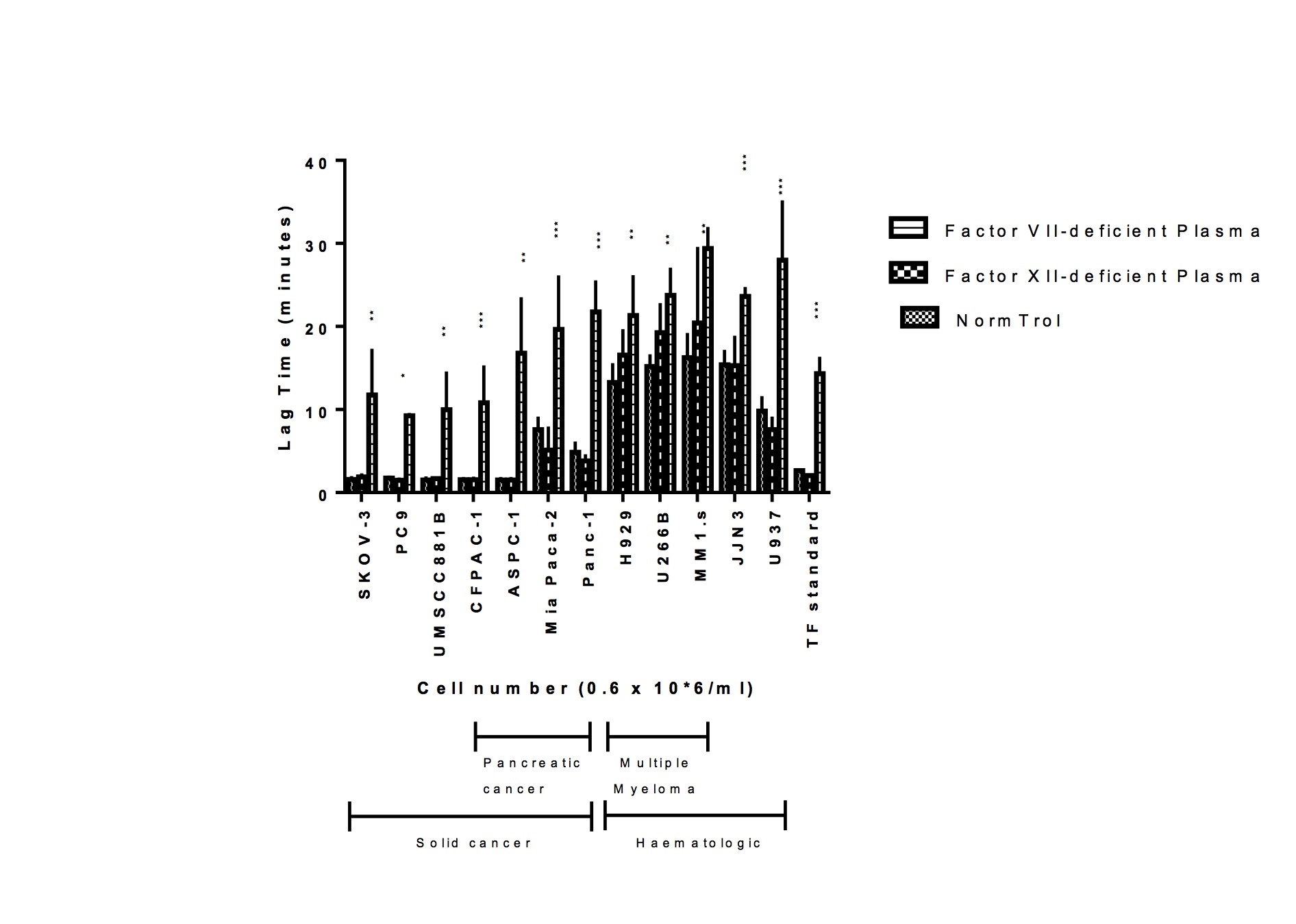
Figure 1. Thrombin generation curves of cancer cells. Thrombograms show TG in NormTrol platelet-free plasma induced by 12 cancer cell lines with low 1pM TF as control.
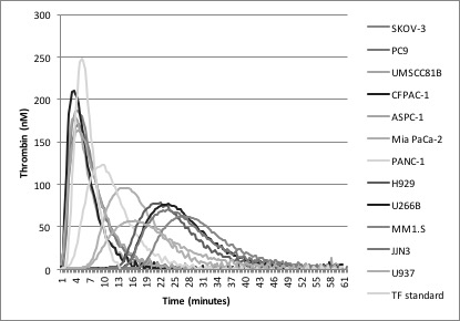
Figure 2: Influence of coagulation factors on thrombin generation in cell lines. TG in solid cancer cell lines was compared with hematologic cell lines in vivo on the CAT assay, at 0.6 x 10*6/ml concentration. Results are mean of n=4 +/- S.D performed in duplicates. *P values from two-way ANOVA are between NormTrol and Factor VII-deficient plasma as those between NormTrol and Factor XII-deficient plasma are mostly insignificant.
Conclusion
This study shows that the specific coagulation factors present in the intrinsic or extrinsic arms of the clotting cascade have a markedly different contribution to the thrombin generation profile of hematologic versus solid malignancies as measured by the CAT assay.
References
1. Hemker HC, Al Dieri R, De Smedt E, Beguin S. Thrombin generation, a function test of the haemostatic-thrombotic system. Thromb Haemost. 2006;96(5):553-61.
Abstract: O06
Type: Oral presentation
Session Topic: Inherited and acquired coagulation disorders
Presentation during EHA Scientific Conference on Bleeding Disorders:
On Thursday, September 15, 2016 from 09:00 - 10:30
Location: Rubí + Zafir
Background
The calibrated automated thrombogram (CAT) assay is emerging as a reliable tool for real time estimation of thrombin generation (TG) potential. There is limited knowledge about the differences in the pathways that underlie the thrombotic phenotype in the solid versus haematological malignant condition. Few studies have investigated the contribution to thrombosis of different factors of the coagulation cascade1. Characterizing the TG capacity in these two distinct cancer models using factor deficient plasma might potentially allow better characterization of the thrombotic risk and individualization of prevention strategies.
Methods
Solid tumour cells (pancreatic cancer AsPC-1, CFPAC-1, PANC-1, MIA PaCa-2 and others such as SKOV-3 ovarian cancer, UMSCC81B Head & Neck squamous cancer, PC9 lung cancer) and malignant haematological cell lines (Multiple myeloma MM1.S, U266B, H929 and others including JJN3 plasma cell leukaemia, U937 histiocytic lymphoma) were evaluated at increasing cell concentrations on a Thrombinoscope for the CAT assay, with the addition of platelet-free normal plasma (NormTrol) or plasma deficient in coagulation factors VII and XII, and TF 1pM standard preparations as control. In addition, tissue factor (TF) cell surface expression was measured with flow cytometry.
Results
In NormTrol plasma, TG in all cancer cell lines was concentration dependent, with CFPAC-1 producing the highest thrombin. Absence of Factor-VII in platelet-free plasma resulted in significantly higher inhibition of TG in solid cancers compared to haematological cancers (Figure 2). TF surface expression correlated strongly with the TG parameters e.g. the higher the TF expressed in a cell line, then the shorter the Lag time and time-to-peak, and UMSCC81B expressed the highest TF. Compared to 1pM TF control, solid tumour cell lines had higher thrombin peaks, faster lag times, and a TG profile of overall greater magnitude than haematological cell lines.
Figure 1. Thrombin generation curves of cancer cells. Thrombograms show TG in NormTrol platelet-free plasma induced by 12 cancer cell lines with low 1pM TF as control.
Figure 2: Influence of coagulation factors on thrombin generation in cell lines. TG in solid cancer cell lines was compared with hematologic cell lines in vivo on the CAT assay, at 0.6 x 10*6/ml concentration. Results are mean of n=4 +/- S.D performed in duplicates. *P values from two-way ANOVA are between NormTrol and Factor VII-deficient plasma as those between NormTrol and Factor XII-deficient plasma are mostly insignificant.
Conclusion
This study shows that the specific coagulation factors present in the intrinsic or extrinsic arms of the clotting cascade have a markedly different contribution to the thrombin generation profile of hematologic versus solid malignancies as measured by the CAT assay.
References
1. Hemker HC, Al Dieri R, De Smedt E, Beguin S. Thrombin generation, a function test of the haemostatic-thrombotic system. Thromb Haemost. 2006;96(5):553-61.