
MICROVASCULAR NETWORK IN BONE MARROWS OF TREATMENT-NAÏVE PATIENTS WITH TYPE 1 GAUCHER DISEASE: SKEWED ANGIOGENIC SIGNALS IN AN INFLAMMATORY MILIEU
(Abstract release date: 05/19/16)
EHA Library. Klimkowska M. 06/09/16; 135375; LB2264

Dr. Monika Klimkowska
Contributions
Contributions
Abstract
Abstract: LB2264
Type: Eposter Presentation
Background
Gaucher disease (GD) is characterized by accumulation of glucocerebroside-storing macrophages, called Gaucher cells (GC) in various organs, including bone marrow (BM). Since macrophages play a major role in chronic inflammation, it can be assumed that macrophage proliferation in GD can elicit or modify local inflammation-associated phenomena, including angiogenesis.
Aims
The study was aim to evaluate microvascular network in BM samples from untreated patients with GD type 1, and compare them to samples from healthy adults.
Methods
Bone marrow core biopsies from 11 treatment-naïve patients with GD type 1 (3 women, 8 men, median age 68.5 years), followed at the Hematology Center, Karolinska University Hospital, were analyzed retrospectively. Biopsies from 36 subjects (18 women, 18 men, median age 63.6 yrs) with no hematological diseases, were used as controls. In the immunohistochemically and immunofluorescent stained samples, the following parameters were analyzed: cellularity, vessel morphology, microvascular density (MVD), vessel wall pericyte coverage and expression of proangiogenic growth factors (VEGF, ANGPT1 and 2). In GD biopsies, analyzes were performed separately in areas with greater and lower percentage of infiltrating GC (≥50% and <50% in a high power field).
Results
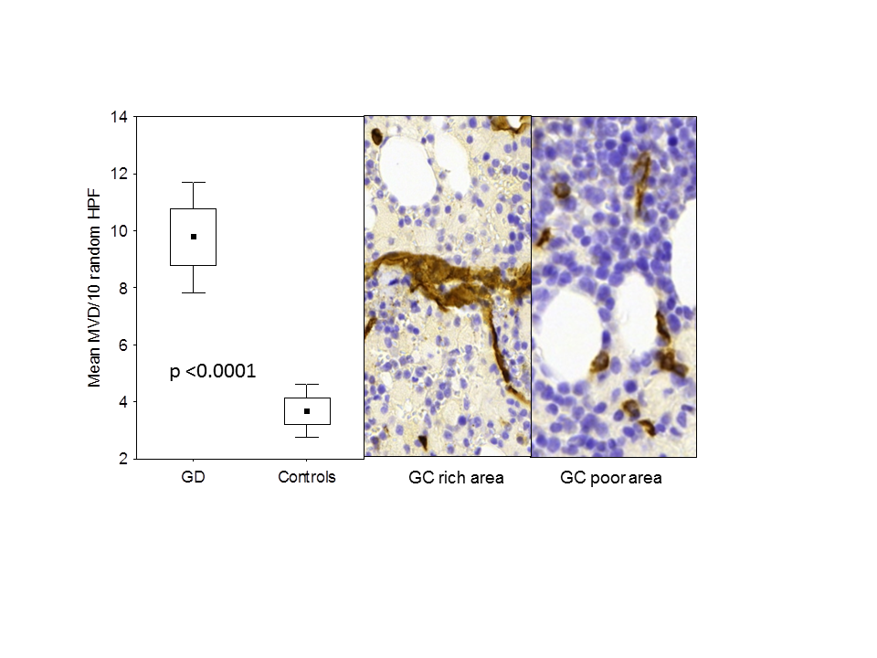
In GD patients, hematopoietic tissue was hypercellular for age, and MVD was 2.6-fold higher than in controls (p<0.001). In GC rich areas, MVD was 1.4-fold higher (p=0.026) and vessel architecture was abnormal as compared to GC poor areas. Moreover, 30 ± 17% of GD BM vessels were pericyte-coated, significantly fewer than in controls (48 ± 16%; p<0.001). Expression of ANGPT1 and 2 was significantly higher in BM vessel walls in GD samples than in controls (7.2 and 13.2 fold higher, respectively), whereas VEGF expression was 20-fold lower in GD (p<0.05 for all parameters). MVD values correlated with BM cellularity, particularly in GC rich areas. Gaucher cells stain moderately for ANGPT1 and 2, and variably for VEGF.
Conclusion
Bone marrow in patients with GD type 1 demonstrates abnormal angiogenesis, with defective pericyte coating, and skewed VEGF/angiopoietin balance, which is presumably related to local accumulation of Gaucher cells.
Session topic: E-poster
Keyword(s): Angiogenesis, Bone Marrow, Gaucher disease
Type: Eposter Presentation
Background
Gaucher disease (GD) is characterized by accumulation of glucocerebroside-storing macrophages, called Gaucher cells (GC) in various organs, including bone marrow (BM). Since macrophages play a major role in chronic inflammation, it can be assumed that macrophage proliferation in GD can elicit or modify local inflammation-associated phenomena, including angiogenesis.
Aims
The study was aim to evaluate microvascular network in BM samples from untreated patients with GD type 1, and compare them to samples from healthy adults.
Methods
Bone marrow core biopsies from 11 treatment-naïve patients with GD type 1 (3 women, 8 men, median age 68.5 years), followed at the Hematology Center, Karolinska University Hospital, were analyzed retrospectively. Biopsies from 36 subjects (18 women, 18 men, median age 63.6 yrs) with no hematological diseases, were used as controls. In the immunohistochemically and immunofluorescent stained samples, the following parameters were analyzed: cellularity, vessel morphology, microvascular density (MVD), vessel wall pericyte coverage and expression of proangiogenic growth factors (VEGF, ANGPT1 and 2). In GD biopsies, analyzes were performed separately in areas with greater and lower percentage of infiltrating GC (≥50% and <50% in a high power field).
Results
In GD patients, hematopoietic tissue was hypercellular for age, and MVD was 2.6-fold higher than in controls (p<0.001). In GC rich areas, MVD was 1.4-fold higher (p=0.026) and vessel architecture was abnormal as compared to GC poor areas. Moreover, 30 ± 17% of GD BM vessels were pericyte-coated, significantly fewer than in controls (48 ± 16%; p<0.001). Expression of ANGPT1 and 2 was significantly higher in BM vessel walls in GD samples than in controls (7.2 and 13.2 fold higher, respectively), whereas VEGF expression was 20-fold lower in GD (p<0.05 for all parameters). MVD values correlated with BM cellularity, particularly in GC rich areas. Gaucher cells stain moderately for ANGPT1 and 2, and variably for VEGF.
Conclusion
Bone marrow in patients with GD type 1 demonstrates abnormal angiogenesis, with defective pericyte coating, and skewed VEGF/angiopoietin balance, which is presumably related to local accumulation of Gaucher cells.
Session topic: E-poster
Keyword(s): Angiogenesis, Bone Marrow, Gaucher disease
Abstract: LB2264
Type: Eposter Presentation
Background
Gaucher disease (GD) is characterized by accumulation of glucocerebroside-storing macrophages, called Gaucher cells (GC) in various organs, including bone marrow (BM). Since macrophages play a major role in chronic inflammation, it can be assumed that macrophage proliferation in GD can elicit or modify local inflammation-associated phenomena, including angiogenesis.
Aims
The study was aim to evaluate microvascular network in BM samples from untreated patients with GD type 1, and compare them to samples from healthy adults.
Methods
Bone marrow core biopsies from 11 treatment-naïve patients with GD type 1 (3 women, 8 men, median age 68.5 years), followed at the Hematology Center, Karolinska University Hospital, were analyzed retrospectively. Biopsies from 36 subjects (18 women, 18 men, median age 63.6 yrs) with no hematological diseases, were used as controls. In the immunohistochemically and immunofluorescent stained samples, the following parameters were analyzed: cellularity, vessel morphology, microvascular density (MVD), vessel wall pericyte coverage and expression of proangiogenic growth factors (VEGF, ANGPT1 and 2). In GD biopsies, analyzes were performed separately in areas with greater and lower percentage of infiltrating GC (≥50% and <50% in a high power field).
Results
In GD patients, hematopoietic tissue was hypercellular for age, and MVD was 2.6-fold higher than in controls (p<0.001). In GC rich areas, MVD was 1.4-fold higher (p=0.026) and vessel architecture was abnormal as compared to GC poor areas. Moreover, 30 ± 17% of GD BM vessels were pericyte-coated, significantly fewer than in controls (48 ± 16%; p<0.001). Expression of ANGPT1 and 2 was significantly higher in BM vessel walls in GD samples than in controls (7.2 and 13.2 fold higher, respectively), whereas VEGF expression was 20-fold lower in GD (p<0.05 for all parameters). MVD values correlated with BM cellularity, particularly in GC rich areas. Gaucher cells stain moderately for ANGPT1 and 2, and variably for VEGF.
Conclusion
Bone marrow in patients with GD type 1 demonstrates abnormal angiogenesis, with defective pericyte coating, and skewed VEGF/angiopoietin balance, which is presumably related to local accumulation of Gaucher cells.
Session topic: E-poster
Keyword(s): Angiogenesis, Bone Marrow, Gaucher disease
{{ help_message }}
{{filter}}