
IDENTIFICATION OF A NOVEL RNA GIANT NUCLEAR BODY IN MYELOID LEUKEMIA
(Abstract release date: 05/19/16)
EHA Library. Zhou H. 06/09/16; 135354; LB2243

Ms. Hong Zhou
Contributions
Contributions
Abstract
Abstract: LB2243
Type: Eposter Presentation
Background
Constitutive synthesis of oncogenic mRNAs is essential for maintaining the uncontrolled growth of myeloid cells. However, little is known about how these mRNAs are exported from the nucleus to the cytoplasm. Cancer cells, unlike normal cells that only divide a finite number of times before they enter into a state of growth arrest or die, are able to maintain uncontrolled proliferation. The cell nucleus, which houses much of the genome and the machinery needed for its replication, maintenance, and expression, is crucial for the survival and proliferation of cells. However, no nuclear bodies have been definitively linked to the uncontrolled proliferation of cancer cells so far. EIF4E nuclear bodies were initially reported two decade ago and thought to be involved in the exporting of nuclear mRNAs associated with cell proliferation. studies have shown a positive correlation between increased eIF4E phosphorylation and cancer cell proliferation as well as tumorigenesis. Consistently, highly phosphorylated eIF4E (p-eIF4E) was frequently observed in a variety of human cancers. Therefore, we hypothesized that cancer-associated nuclear bodies as detected by p-eIF4E antibody may exist in the nuclei of cancer cells.
Aims
To identify the putative cancer-associated nuclear body, we initially stained two human leukemia cell lines KG-1 (acute myeloid leukemia, AML) by immunofluorescence staining with antibodies against phosphorylated eIF4E (p-eIF4E) or total eIF4E (t-eIF4E); To reveal the morphology and structure features of GNB; To determine whether RNA-GNB abundance was associated with the hyperproliferative phenotype in human cancer.
Methods
Cell lines and cultureMyeloid cell lines were used in this study.Hematopoietic malignant cells were cultured in RPMI-1640 supplemented with 10% fetal calf serum (FCS) at 37°C in a 95% air, 5% CO2 umidified incubator. Normal human blood samples and human leukemia cell samplesNormal blood cells and primary leukemia cell samples were isolated from healthy volunteers or leukemia patients with their informed consent in accordance with the Declaration of Helsinki. All experiments were approved by the ethics committee of Hangzhou First People's Hospital.Immunofluorescence stainingCells were fixed with freshly prepared 3.7% paraformaldehyde in PBS (pH 7.2) for 20 min at room temperature on slides. Cells were then blocked and permeabilized with PBS containing 10% FBS and 0.1% Tween-20 for 30 min at room temperature. Staining of cells with primary antibodies was performed overnight at 4°C, and then with a FITC or rhodamine-conjugated secondary antibodies for 1 h at room temperature. After three washes with PBS, the slides were mounted in Vectashield with DAPI and sealed.Immunopurification of GNBs from leukemia cellsGNBs were purified from leukemia cells as centrifugation at 2,000 rpm for 5 minutes at 4°C. After washed three times with PBS, cells were resuspended in PBS containing protease inhibitors and transferred to a 7 mL Dounce tissue homogenizer for dounce homogenization. The homogenized suspension was collected for removing intact cells and undisrupted nuclei by centrifugation at 1,000 rpm for 5 minutes at 4°C. Separation of GNBs from nucleoli on the cell nuclei Cells were collected by centrifugation at 2500 rpm for 5 minutes at 4°C. After removing the supernatant, the cell pellet was lysated with 0.5ml of buffer A for 5min and then centrifuged at 700 rpm, 5min at 4°C. The supernatant was carefully removed and the nuclei were washed with 1.5ml of Buffer A without NP40 by centrifugation at 700 rpm, 5min at 4°C.
Results
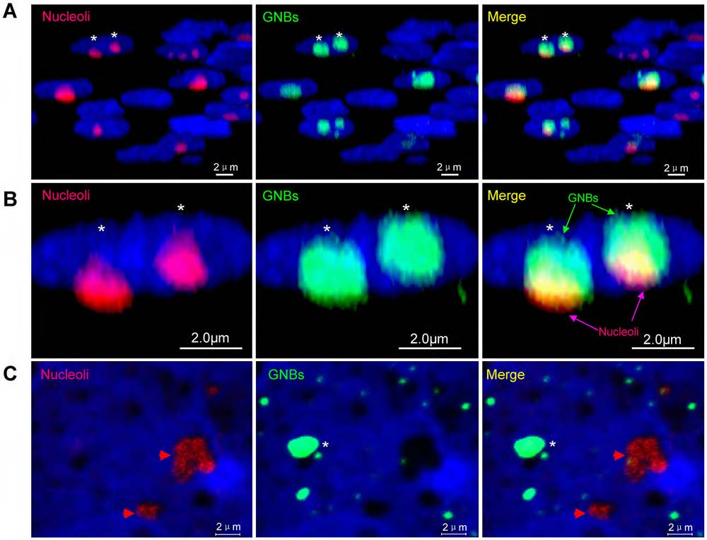
we report the identification of a RNA giant nuclear body (RNA-GNB) that is abundant in acute myeloid cells but rare in normal cells. The RNA-GNB contains a RNA core surrounded by a protein shell. We identify 782 proteins from acute myeloid-associated RNA-GNBs, 40% of which are involved in the nuclear mRNA trafficking. RNA-GNB is required for cell proliferation, and its abundance is positively associated with tumor burden and outcome of therapies.
Conclusion
our studies have identified RNA-GNB as a potential nuclear mRNA trafficking organelle. Further studies will be required to characterize the components of proteins and mRNAs in RNA-GNB and reveal their functions. This will help elucidate how RNA-GNBs form and regulate the uncontrolled proliferation of cancer cells.
Session topic: E-poster
Keyword(s): Cell Trafficking, Myeloid leukemia, Phosphorylation, Proliferation
Type: Eposter Presentation
Background
Constitutive synthesis of oncogenic mRNAs is essential for maintaining the uncontrolled growth of myeloid cells. However, little is known about how these mRNAs are exported from the nucleus to the cytoplasm. Cancer cells, unlike normal cells that only divide a finite number of times before they enter into a state of growth arrest or die, are able to maintain uncontrolled proliferation. The cell nucleus, which houses much of the genome and the machinery needed for its replication, maintenance, and expression, is crucial for the survival and proliferation of cells. However, no nuclear bodies have been definitively linked to the uncontrolled proliferation of cancer cells so far. EIF4E nuclear bodies were initially reported two decade ago and thought to be involved in the exporting of nuclear mRNAs associated with cell proliferation. studies have shown a positive correlation between increased eIF4E phosphorylation and cancer cell proliferation as well as tumorigenesis. Consistently, highly phosphorylated eIF4E (p-eIF4E) was frequently observed in a variety of human cancers. Therefore, we hypothesized that cancer-associated nuclear bodies as detected by p-eIF4E antibody may exist in the nuclei of cancer cells.
Aims
To identify the putative cancer-associated nuclear body, we initially stained two human leukemia cell lines KG-1 (acute myeloid leukemia, AML) by immunofluorescence staining with antibodies against phosphorylated eIF4E (p-eIF4E) or total eIF4E (t-eIF4E); To reveal the morphology and structure features of GNB; To determine whether RNA-GNB abundance was associated with the hyperproliferative phenotype in human cancer.
Methods
Cell lines and cultureMyeloid cell lines were used in this study.Hematopoietic malignant cells were cultured in RPMI-1640 supplemented with 10% fetal calf serum (FCS) at 37°C in a 95% air, 5% CO2 umidified incubator. Normal human blood samples and human leukemia cell samplesNormal blood cells and primary leukemia cell samples were isolated from healthy volunteers or leukemia patients with their informed consent in accordance with the Declaration of Helsinki. All experiments were approved by the ethics committee of Hangzhou First People's Hospital.Immunofluorescence stainingCells were fixed with freshly prepared 3.7% paraformaldehyde in PBS (pH 7.2) for 20 min at room temperature on slides. Cells were then blocked and permeabilized with PBS containing 10% FBS and 0.1% Tween-20 for 30 min at room temperature. Staining of cells with primary antibodies was performed overnight at 4°C, and then with a FITC or rhodamine-conjugated secondary antibodies for 1 h at room temperature. After three washes with PBS, the slides were mounted in Vectashield with DAPI and sealed.Immunopurification of GNBs from leukemia cellsGNBs were purified from leukemia cells as centrifugation at 2,000 rpm for 5 minutes at 4°C. After washed three times with PBS, cells were resuspended in PBS containing protease inhibitors and transferred to a 7 mL Dounce tissue homogenizer for dounce homogenization. The homogenized suspension was collected for removing intact cells and undisrupted nuclei by centrifugation at 1,000 rpm for 5 minutes at 4°C. Separation of GNBs from nucleoli on the cell nuclei Cells were collected by centrifugation at 2500 rpm for 5 minutes at 4°C. After removing the supernatant, the cell pellet was lysated with 0.5ml of buffer A for 5min and then centrifuged at 700 rpm, 5min at 4°C. The supernatant was carefully removed and the nuclei were washed with 1.5ml of Buffer A without NP40 by centrifugation at 700 rpm, 5min at 4°C.
Results
we report the identification of a RNA giant nuclear body (RNA-GNB) that is abundant in acute myeloid cells but rare in normal cells. The RNA-GNB contains a RNA core surrounded by a protein shell. We identify 782 proteins from acute myeloid-associated RNA-GNBs, 40% of which are involved in the nuclear mRNA trafficking. RNA-GNB is required for cell proliferation, and its abundance is positively associated with tumor burden and outcome of therapies.
Conclusion
our studies have identified RNA-GNB as a potential nuclear mRNA trafficking organelle. Further studies will be required to characterize the components of proteins and mRNAs in RNA-GNB and reveal their functions. This will help elucidate how RNA-GNBs form and regulate the uncontrolled proliferation of cancer cells.
Session topic: E-poster
Keyword(s): Cell Trafficking, Myeloid leukemia, Phosphorylation, Proliferation
Abstract: LB2243
Type: Eposter Presentation
Background
Constitutive synthesis of oncogenic mRNAs is essential for maintaining the uncontrolled growth of myeloid cells. However, little is known about how these mRNAs are exported from the nucleus to the cytoplasm. Cancer cells, unlike normal cells that only divide a finite number of times before they enter into a state of growth arrest or die, are able to maintain uncontrolled proliferation. The cell nucleus, which houses much of the genome and the machinery needed for its replication, maintenance, and expression, is crucial for the survival and proliferation of cells. However, no nuclear bodies have been definitively linked to the uncontrolled proliferation of cancer cells so far. EIF4E nuclear bodies were initially reported two decade ago and thought to be involved in the exporting of nuclear mRNAs associated with cell proliferation. studies have shown a positive correlation between increased eIF4E phosphorylation and cancer cell proliferation as well as tumorigenesis. Consistently, highly phosphorylated eIF4E (p-eIF4E) was frequently observed in a variety of human cancers. Therefore, we hypothesized that cancer-associated nuclear bodies as detected by p-eIF4E antibody may exist in the nuclei of cancer cells.
Aims
To identify the putative cancer-associated nuclear body, we initially stained two human leukemia cell lines KG-1 (acute myeloid leukemia, AML) by immunofluorescence staining with antibodies against phosphorylated eIF4E (p-eIF4E) or total eIF4E (t-eIF4E); To reveal the morphology and structure features of GNB; To determine whether RNA-GNB abundance was associated with the hyperproliferative phenotype in human cancer.
Methods
Cell lines and cultureMyeloid cell lines were used in this study.Hematopoietic malignant cells were cultured in RPMI-1640 supplemented with 10% fetal calf serum (FCS) at 37°C in a 95% air, 5% CO2 umidified incubator. Normal human blood samples and human leukemia cell samplesNormal blood cells and primary leukemia cell samples were isolated from healthy volunteers or leukemia patients with their informed consent in accordance with the Declaration of Helsinki. All experiments were approved by the ethics committee of Hangzhou First People's Hospital.Immunofluorescence stainingCells were fixed with freshly prepared 3.7% paraformaldehyde in PBS (pH 7.2) for 20 min at room temperature on slides. Cells were then blocked and permeabilized with PBS containing 10% FBS and 0.1% Tween-20 for 30 min at room temperature. Staining of cells with primary antibodies was performed overnight at 4°C, and then with a FITC or rhodamine-conjugated secondary antibodies for 1 h at room temperature. After three washes with PBS, the slides were mounted in Vectashield with DAPI and sealed.Immunopurification of GNBs from leukemia cellsGNBs were purified from leukemia cells as centrifugation at 2,000 rpm for 5 minutes at 4°C. After washed three times with PBS, cells were resuspended in PBS containing protease inhibitors and transferred to a 7 mL Dounce tissue homogenizer for dounce homogenization. The homogenized suspension was collected for removing intact cells and undisrupted nuclei by centrifugation at 1,000 rpm for 5 minutes at 4°C. Separation of GNBs from nucleoli on the cell nuclei Cells were collected by centrifugation at 2500 rpm for 5 minutes at 4°C. After removing the supernatant, the cell pellet was lysated with 0.5ml of buffer A for 5min and then centrifuged at 700 rpm, 5min at 4°C. The supernatant was carefully removed and the nuclei were washed with 1.5ml of Buffer A without NP40 by centrifugation at 700 rpm, 5min at 4°C.
Results
we report the identification of a RNA giant nuclear body (RNA-GNB) that is abundant in acute myeloid cells but rare in normal cells. The RNA-GNB contains a RNA core surrounded by a protein shell. We identify 782 proteins from acute myeloid-associated RNA-GNBs, 40% of which are involved in the nuclear mRNA trafficking. RNA-GNB is required for cell proliferation, and its abundance is positively associated with tumor burden and outcome of therapies.
Conclusion
our studies have identified RNA-GNB as a potential nuclear mRNA trafficking organelle. Further studies will be required to characterize the components of proteins and mRNAs in RNA-GNB and reveal their functions. This will help elucidate how RNA-GNBs form and regulate the uncontrolled proliferation of cancer cells.
Session topic: E-poster
Keyword(s): Cell Trafficking, Myeloid leukemia, Phosphorylation, Proliferation
{{ help_message }}
{{filter}}