
MAGIC-TT-MEDIATED CELLS TARGET TRANSPLANTATION INTO BONE MARROW WHICH INCLUDES THE STUDY OF CELL DISTRIBUTION IN VIVO AND THE EFFECTS OF HEMATOPOIETIC RECONSTRUCTION.
(Abstract release date: 05/19/16)
EHA Library. Huang H. 06/09/16; 134746; PB1846

Dr. Hao Huang
Contributions
Contributions
Abstract
Abstract: PB1846
Type: Publication Only
Background
Target cell transplantation to bone marrow (BM) is possible by Magnetism-induced cell target transplantation (MagiC-TT) (EHA2015posterP701).
Aims
RFP-MSC were used to observe the cell’s distribution and the following effect, additionally CD45+ GFP cells were used to evaluate the promotion of hematopoietic reconstitution of MagiC-TT.
Methods
1)Target transplantation of MSC: Fe3O4@PDA@Au nanoparticles (NPs) were synthesized and introduced into Luciferase gene modified RFP-MSC (Luc-RFP-MSCs). Magnetized and wt Luc-RFP-MSCs were compared on the biological feature and their ability of migration ex vivo. Then magnetized RFP-MSCs were micro-injected into the femur cavity of the mice with the help of X-ray (Fig.1a), under magnet in M group or without in W group. Bioluminescence, FACS, PCR and histopathological analysis were ued after transplantation. 20 eGFP transgenic mice and another 20 C57 mice were used. 2) CD45+ cells transplantation: 34 C57 mice were randomly divided into 2 groups evenly. 7.5Gy myeloablative irradiation were given 1d before, then CD45+ cells were isolated by MACS from eGFP transgenic mice and freshly micro-injected into right femur, 1×10⁶ cells/20uL per mouse, with or without magnet for 24hrs (M or W group). At 0h, 24h, 72h after injection, every 3 mice in both groups were sacrificed at each time point for detection, then remaining 8 mice in both groups were compared with the general conditions, hematopoietic recovery, GFP+ cells in different organs etc.
Results
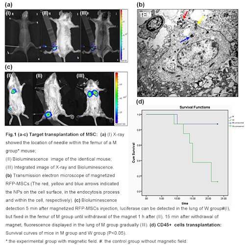
1) Target transplantation of MSCs: NPs exist within or on the surface of magnetized RFP-MSCs (Fig.1b), no obvious change was found. The magnetized RFP-MSCs were capable of target migration under magnetism ex vivo. Bioluminescence assay showed that the magnetized Luc-RFP-MSCs appeared in the lung of W group 5 min after cell injection, while fixed in the femur of M group mice. However, on withdrawal of magnet 1h after cell injection, strong fluorescence was observed in the lung of M group gradually (Fig.1c). By pathological examinations, FACS and PCR, large number of RFP-MSCs were observed to reside within the BM in M group while few in W group, thereby demonstrating the specific BM target transplantation of magnetized RFP-MSCs. Those RFP-MSCs were found to survive more than 3m in different organs. 2) CD45+ cells transplantation: The mice that survived were 7/8 vs 1/8 in M or W group respectively (Fig.1d). The GFP% in femurs of both groups (Tab.1) also proved the specific BM target transplantation in M group. Platelet recovery in M group is faster than that in W group (12.33d±2.42d vs 16.38d±2.39d, P=0.009); and the lowest value of decreased hemoglobin in M group was higher than that of W group (43.75±13.02 vs 13.75±5.18, P<0.001).
Conclusion
Fe3O4@PDA@Au NPs rendered MSCs their capability of target transplantation by MagiC-TT technique. MagiC-TT also helps in the recovery of PLT after transplantation after CD45+ hematopoietic cell transplantation .
Session topic: E-poster
Keyword(s): GFP, HSCT, Nanoparticle, Targeted therapy
Type: Publication Only
Background
Target cell transplantation to bone marrow (BM) is possible by Magnetism-induced cell target transplantation (MagiC-TT) (EHA2015posterP701).
Aims
RFP-MSC were used to observe the cell’s distribution and the following effect, additionally CD45+ GFP cells were used to evaluate the promotion of hematopoietic reconstitution of MagiC-TT.
Methods
1)Target transplantation of MSC: Fe3O4@PDA@Au nanoparticles (NPs) were synthesized and introduced into Luciferase gene modified RFP-MSC (Luc-RFP-MSCs). Magnetized and wt Luc-RFP-MSCs were compared on the biological feature and their ability of migration ex vivo. Then magnetized RFP-MSCs were micro-injected into the femur cavity of the mice with the help of X-ray (Fig.1a), under magnet in M group or without in W group. Bioluminescence, FACS, PCR and histopathological analysis were ued after transplantation. 20 eGFP transgenic mice and another 20 C57 mice were used. 2) CD45+ cells transplantation: 34 C57 mice were randomly divided into 2 groups evenly. 7.5Gy myeloablative irradiation were given 1d before, then CD45+ cells were isolated by MACS from eGFP transgenic mice and freshly micro-injected into right femur, 1×10⁶ cells/20uL per mouse, with or without magnet for 24hrs (M or W group). At 0h, 24h, 72h after injection, every 3 mice in both groups were sacrificed at each time point for detection, then remaining 8 mice in both groups were compared with the general conditions, hematopoietic recovery, GFP+ cells in different organs etc.
Results
1) Target transplantation of MSCs: NPs exist within or on the surface of magnetized RFP-MSCs (Fig.1b), no obvious change was found. The magnetized RFP-MSCs were capable of target migration under magnetism ex vivo. Bioluminescence assay showed that the magnetized Luc-RFP-MSCs appeared in the lung of W group 5 min after cell injection, while fixed in the femur of M group mice. However, on withdrawal of magnet 1h after cell injection, strong fluorescence was observed in the lung of M group gradually (Fig.1c). By pathological examinations, FACS and PCR, large number of RFP-MSCs were observed to reside within the BM in M group while few in W group, thereby demonstrating the specific BM target transplantation of magnetized RFP-MSCs. Those RFP-MSCs were found to survive more than 3m in different organs. 2) CD45+ cells transplantation: The mice that survived were 7/8 vs 1/8 in M or W group respectively (Fig.1d). The GFP% in femurs of both groups (Tab.1) also proved the specific BM target transplantation in M group. Platelet recovery in M group is faster than that in W group (12.33d±2.42d vs 16.38d±2.39d, P=0.009); and the lowest value of decreased hemoglobin in M group was higher than that of W group (43.75±13.02 vs 13.75±5.18, P<0.001).
| Table 1. Comparison of the percentage of RFP-MSCs in M group and W group by flow cytometry. | |||||||||
| group | 0h(%) | p | 24h(%) | p | 72h(%) | p | |||
| *LC | **RT | LC | RT | LC | RT | ||||
| BMM | 0.017±0.006 | 0.497±0.151 | 0.040 | 0.080±0.026 | 1.573±0.508 | 0.030 | 0.190±0.139 | 1.960±0.809 | 0.049 |
| BMW | 0.017±0.012 | 0.050±0.017 | 0.184 | 0.013±0.006 | 0.027±0.015 | 0.184 | 0.023±0.015 | 0.320±0.434 | 0.368 |
| P | 1.000 | 0.007 | 0.013 | 0.006 | 0.108 | 0.036 | |||
| *:Left control femur; **: Right experimental femur. | |||||||||
Conclusion
Fe3O4@PDA@Au NPs rendered MSCs their capability of target transplantation by MagiC-TT technique. MagiC-TT also helps in the recovery of PLT after transplantation after CD45+ hematopoietic cell transplantation .
Session topic: E-poster
Keyword(s): GFP, HSCT, Nanoparticle, Targeted therapy
Abstract: PB1846
Type: Publication Only
Background
Target cell transplantation to bone marrow (BM) is possible by Magnetism-induced cell target transplantation (MagiC-TT) (EHA2015posterP701).
Aims
RFP-MSC were used to observe the cell’s distribution and the following effect, additionally CD45+ GFP cells were used to evaluate the promotion of hematopoietic reconstitution of MagiC-TT.
Methods
1)Target transplantation of MSC: Fe3O4@PDA@Au nanoparticles (NPs) were synthesized and introduced into Luciferase gene modified RFP-MSC (Luc-RFP-MSCs). Magnetized and wt Luc-RFP-MSCs were compared on the biological feature and their ability of migration ex vivo. Then magnetized RFP-MSCs were micro-injected into the femur cavity of the mice with the help of X-ray (Fig.1a), under magnet in M group or without in W group. Bioluminescence, FACS, PCR and histopathological analysis were ued after transplantation. 20 eGFP transgenic mice and another 20 C57 mice were used. 2) CD45+ cells transplantation: 34 C57 mice were randomly divided into 2 groups evenly. 7.5Gy myeloablative irradiation were given 1d before, then CD45+ cells were isolated by MACS from eGFP transgenic mice and freshly micro-injected into right femur, 1×10⁶ cells/20uL per mouse, with or without magnet for 24hrs (M or W group). At 0h, 24h, 72h after injection, every 3 mice in both groups were sacrificed at each time point for detection, then remaining 8 mice in both groups were compared with the general conditions, hematopoietic recovery, GFP+ cells in different organs etc.
Results
1) Target transplantation of MSCs: NPs exist within or on the surface of magnetized RFP-MSCs (Fig.1b), no obvious change was found. The magnetized RFP-MSCs were capable of target migration under magnetism ex vivo. Bioluminescence assay showed that the magnetized Luc-RFP-MSCs appeared in the lung of W group 5 min after cell injection, while fixed in the femur of M group mice. However, on withdrawal of magnet 1h after cell injection, strong fluorescence was observed in the lung of M group gradually (Fig.1c). By pathological examinations, FACS and PCR, large number of RFP-MSCs were observed to reside within the BM in M group while few in W group, thereby demonstrating the specific BM target transplantation of magnetized RFP-MSCs. Those RFP-MSCs were found to survive more than 3m in different organs. 2) CD45+ cells transplantation: The mice that survived were 7/8 vs 1/8 in M or W group respectively (Fig.1d). The GFP% in femurs of both groups (Tab.1) also proved the specific BM target transplantation in M group. Platelet recovery in M group is faster than that in W group (12.33d±2.42d vs 16.38d±2.39d, P=0.009); and the lowest value of decreased hemoglobin in M group was higher than that of W group (43.75±13.02 vs 13.75±5.18, P<0.001).
Conclusion
Fe3O4@PDA@Au NPs rendered MSCs their capability of target transplantation by MagiC-TT technique. MagiC-TT also helps in the recovery of PLT after transplantation after CD45+ hematopoietic cell transplantation .
Session topic: E-poster
Keyword(s): GFP, HSCT, Nanoparticle, Targeted therapy
Type: Publication Only
Background
Target cell transplantation to bone marrow (BM) is possible by Magnetism-induced cell target transplantation (MagiC-TT) (EHA2015posterP701).
Aims
RFP-MSC were used to observe the cell’s distribution and the following effect, additionally CD45+ GFP cells were used to evaluate the promotion of hematopoietic reconstitution of MagiC-TT.
Methods
1)Target transplantation of MSC: Fe3O4@PDA@Au nanoparticles (NPs) were synthesized and introduced into Luciferase gene modified RFP-MSC (Luc-RFP-MSCs). Magnetized and wt Luc-RFP-MSCs were compared on the biological feature and their ability of migration ex vivo. Then magnetized RFP-MSCs were micro-injected into the femur cavity of the mice with the help of X-ray (Fig.1a), under magnet in M group or without in W group. Bioluminescence, FACS, PCR and histopathological analysis were ued after transplantation. 20 eGFP transgenic mice and another 20 C57 mice were used. 2) CD45+ cells transplantation: 34 C57 mice were randomly divided into 2 groups evenly. 7.5Gy myeloablative irradiation were given 1d before, then CD45+ cells were isolated by MACS from eGFP transgenic mice and freshly micro-injected into right femur, 1×10⁶ cells/20uL per mouse, with or without magnet for 24hrs (M or W group). At 0h, 24h, 72h after injection, every 3 mice in both groups were sacrificed at each time point for detection, then remaining 8 mice in both groups were compared with the general conditions, hematopoietic recovery, GFP+ cells in different organs etc.
Results
1) Target transplantation of MSCs: NPs exist within or on the surface of magnetized RFP-MSCs (Fig.1b), no obvious change was found. The magnetized RFP-MSCs were capable of target migration under magnetism ex vivo. Bioluminescence assay showed that the magnetized Luc-RFP-MSCs appeared in the lung of W group 5 min after cell injection, while fixed in the femur of M group mice. However, on withdrawal of magnet 1h after cell injection, strong fluorescence was observed in the lung of M group gradually (Fig.1c). By pathological examinations, FACS and PCR, large number of RFP-MSCs were observed to reside within the BM in M group while few in W group, thereby demonstrating the specific BM target transplantation of magnetized RFP-MSCs. Those RFP-MSCs were found to survive more than 3m in different organs. 2) CD45+ cells transplantation: The mice that survived were 7/8 vs 1/8 in M or W group respectively (Fig.1d). The GFP% in femurs of both groups (Tab.1) also proved the specific BM target transplantation in M group. Platelet recovery in M group is faster than that in W group (12.33d±2.42d vs 16.38d±2.39d, P=0.009); and the lowest value of decreased hemoglobin in M group was higher than that of W group (43.75±13.02 vs 13.75±5.18, P<0.001).
| Table 1. Comparison of the percentage of RFP-MSCs in M group and W group by flow cytometry. | |||||||||
| group | 0h(%) | p | 24h(%) | p | 72h(%) | p | |||
| *LC | **RT | LC | RT | LC | RT | ||||
| BMM | 0.017±0.006 | 0.497±0.151 | 0.040 | 0.080±0.026 | 1.573±0.508 | 0.030 | 0.190±0.139 | 1.960±0.809 | 0.049 |
| BMW | 0.017±0.012 | 0.050±0.017 | 0.184 | 0.013±0.006 | 0.027±0.015 | 0.184 | 0.023±0.015 | 0.320±0.434 | 0.368 |
| P | 1.000 | 0.007 | 0.013 | 0.006 | 0.108 | 0.036 | |||
| *:Left control femur; **: Right experimental femur. | |||||||||
Conclusion
Fe3O4@PDA@Au NPs rendered MSCs their capability of target transplantation by MagiC-TT technique. MagiC-TT also helps in the recovery of PLT after transplantation after CD45+ hematopoietic cell transplantation .
Session topic: E-poster
Keyword(s): GFP, HSCT, Nanoparticle, Targeted therapy
{{ help_message }}
{{filter}}