
CD34 POSITIVE CELL COUNT DETECTED BY MULTI-COLOR FLOW CYTOMETRY (MFC) AFTER FIRST INDUCTION THERAPY IN ACUTE MYELOID LEUKEMIA (AML) EFFECTS ON SURVIVAL AND RELAPSE
(Abstract release date: 05/19/16)
EHA Library. Atilla E. 06/09/16; 134570; PB1670

Dr. Erden Atilla
Contributions
Contributions
Abstract
Abstract: PB1670
Type: Publication Only
Background
Minimal residual disease (MRD) is defined as detection of low levels of leukemic cells and had been shown to correlate with an increased risk of relapse and shortened survival in Acute Lymphoblastic Leukemia. However there is a controversy in definition of MRD in AML.
Aims
Our aim is to evaluate the effects of CD34 positive cell count of >1.0% after first induction therapy on survival and relapse in AML in our cohort.
Methods
CD34 positive cell count of >1.0% was measured in newly diagnosed 148 AML (excluding acute promyelocytic leukemia) patients by multi-color flow-cytometry (MFC) based detection after first induction regimen in between 2007 and 2015. Chi-square test and kaplan-meier curves were used for statistical analysis. P<0.05 was considered statistically significant.
Results
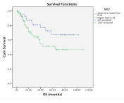
The median age was 47 (range, 21-82). 78 (52%) of patients were male. AML risk stratification at diagnosis were as follows; low risk (21%), standart risk (38%), high risk (22%). For the induction regimen, 11 geriatric patients received azacytidine and 137 patients got standart 3+7 (ARA-C/daunorubicin). 23.5% of patients (35/148) were not in remission and received reinduction therapy. CD34 positive cell count>1.0% was detected in 49.7% patients (74/148) after induction. High dose ARA-C consolidation treatments (mean 2 cycles, range 1-6) were administered and 87 patients (58%) underwent allogeneic-stem cell transplantation due to their risk status. The relapse rate of patients with CD34 positive cell count >1.0% was significantly higher than CD34 positive cell count lower than 1.0% (58% vs 32%, P=0.01). RFS rate at 12 months was higher in CD34 positive cell count lower than 1.0% (72% vs 41%, P=0.1). 2 year OS significantly improved in patients with in CD34 positive cell count lower than 1.0% (83% vs 66%, P=0.049) (Fig)
Conclusion
CD34 positive cell count >1.0% detected by MFC after first induction is related to higher risk of relapse and decreaced OS. Despite techniques for detection of MRD in AML evolve rapidly, MFC remains a useful tool.
Session topic: E-poster
Keyword(s): Acute myeloid leukemia
Type: Publication Only
Background
Minimal residual disease (MRD) is defined as detection of low levels of leukemic cells and had been shown to correlate with an increased risk of relapse and shortened survival in Acute Lymphoblastic Leukemia. However there is a controversy in definition of MRD in AML.
Aims
Our aim is to evaluate the effects of CD34 positive cell count of >1.0% after first induction therapy on survival and relapse in AML in our cohort.
Methods
CD34 positive cell count of >1.0% was measured in newly diagnosed 148 AML (excluding acute promyelocytic leukemia) patients by multi-color flow-cytometry (MFC) based detection after first induction regimen in between 2007 and 2015. Chi-square test and kaplan-meier curves were used for statistical analysis. P<0.05 was considered statistically significant.
Results
The median age was 47 (range, 21-82). 78 (52%) of patients were male. AML risk stratification at diagnosis were as follows; low risk (21%), standart risk (38%), high risk (22%). For the induction regimen, 11 geriatric patients received azacytidine and 137 patients got standart 3+7 (ARA-C/daunorubicin). 23.5% of patients (35/148) were not in remission and received reinduction therapy. CD34 positive cell count>1.0% was detected in 49.7% patients (74/148) after induction. High dose ARA-C consolidation treatments (mean 2 cycles, range 1-6) were administered and 87 patients (58%) underwent allogeneic-stem cell transplantation due to their risk status. The relapse rate of patients with CD34 positive cell count >1.0% was significantly higher than CD34 positive cell count lower than 1.0% (58% vs 32%, P=0.01). RFS rate at 12 months was higher in CD34 positive cell count lower than 1.0% (72% vs 41%, P=0.1). 2 year OS significantly improved in patients with in CD34 positive cell count lower than 1.0% (83% vs 66%, P=0.049) (Fig)
Conclusion
CD34 positive cell count >1.0% detected by MFC after first induction is related to higher risk of relapse and decreaced OS. Despite techniques for detection of MRD in AML evolve rapidly, MFC remains a useful tool.
Session topic: E-poster
Keyword(s): Acute myeloid leukemia
Abstract: PB1670
Type: Publication Only
Background
Minimal residual disease (MRD) is defined as detection of low levels of leukemic cells and had been shown to correlate with an increased risk of relapse and shortened survival in Acute Lymphoblastic Leukemia. However there is a controversy in definition of MRD in AML.
Aims
Our aim is to evaluate the effects of CD34 positive cell count of >1.0% after first induction therapy on survival and relapse in AML in our cohort.
Methods
CD34 positive cell count of >1.0% was measured in newly diagnosed 148 AML (excluding acute promyelocytic leukemia) patients by multi-color flow-cytometry (MFC) based detection after first induction regimen in between 2007 and 2015. Chi-square test and kaplan-meier curves were used for statistical analysis. P<0.05 was considered statistically significant.
Results
The median age was 47 (range, 21-82). 78 (52%) of patients were male. AML risk stratification at diagnosis were as follows; low risk (21%), standart risk (38%), high risk (22%). For the induction regimen, 11 geriatric patients received azacytidine and 137 patients got standart 3+7 (ARA-C/daunorubicin). 23.5% of patients (35/148) were not in remission and received reinduction therapy. CD34 positive cell count>1.0% was detected in 49.7% patients (74/148) after induction. High dose ARA-C consolidation treatments (mean 2 cycles, range 1-6) were administered and 87 patients (58%) underwent allogeneic-stem cell transplantation due to their risk status. The relapse rate of patients with CD34 positive cell count >1.0% was significantly higher than CD34 positive cell count lower than 1.0% (58% vs 32%, P=0.01). RFS rate at 12 months was higher in CD34 positive cell count lower than 1.0% (72% vs 41%, P=0.1). 2 year OS significantly improved in patients with in CD34 positive cell count lower than 1.0% (83% vs 66%, P=0.049) (Fig)
Conclusion
CD34 positive cell count >1.0% detected by MFC after first induction is related to higher risk of relapse and decreaced OS. Despite techniques for detection of MRD in AML evolve rapidly, MFC remains a useful tool.
Session topic: E-poster
Keyword(s): Acute myeloid leukemia
{{ help_message }}
{{filter}}