
MINIMAL RESIDUAL DISEASE: COMPARISON BETWEEN TWO METHODS IN A CLINICAL DIAGNOSTIC LABORATORY.
(Abstract release date: 05/19/16)
EHA Library. Lemes Castellano A. 06/09/16; 134511; PB1611

Angelina Lemes Castellano
Contributions
Contributions
Abstract
Abstract: PB1611
Type: Publication Only
Background
Flow cytometry and molecular techniques are the most important methods for monitoring minimal residual disease (MRD) in leukemia.Evaluation of specific molecular markers represents the most sensitive and specific analysis in MRD leukemia, however the existence a small number of markers which in few cases are valid to follow the course of the disease is a limiting factor. Because of this, there is the necessity to identify new widely represented markers in AML and that could predict relapseOn the other hand, immunophenotype associated to leukemia (LAIP) have been proved to be very useful in the detection of MRD and it can be used up to 85% of patients.
Aims
The aim of this study was to compare the concordance of MRD measured by flow cytometry with standardized molecular techniques including NPM1, Inv16 and AML1-ETO. Furthermore, we also analyzed the behavior of WT1 at this respect in order to determine whether overexpression of WT1 could be a new MRD marker.
Methods
A total of 35 samples of bone marrow from patients with AML in different stages of the disease were assessed. We analyzed NPM1, Inv16 and AML1-ETO by using the gold standard quantitative PCR techniques and these results were compared first with the results obtained by flow cytometry, and then with WT1 levels measured through non standardized quantitative PCR.In order to analyze antigen expression by flow cytometry, we used a combination of eight monoclonal antibodies, according to the panels and procedures suggested by EuroFlow Group. We analyzed about 1,000,000 events to asses MRD with a sensitivity of 10-4, so MRD was registered as positive when we detected at least 100 events with LAIP, (MRD= 0.01%).
Results
Our results revealed:
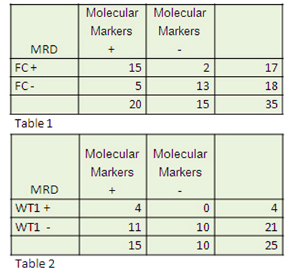
- High concordance between MRD by Flow Cytometry and standardized molecular techniques, with a sensitivity of 88% and a specificity of 72% (Table 1)
- However, when we compared standardized vs non standardized techniques, WT1 showed a sensitivity of 100% and specificity of 47% (Table 2).
Conclusion
1. Flow cytometry is a useful tool for detecting MRD in AML. However, it is important to establish cutoffs to predict relapse in the different stages of the disease.
2. The results regarding WT1 may reflect the lack of standardization in the quantification of the marker, although it shows high specificity for the presence of disease.
Session topic: E-poster
Keyword(s): Flow cytometry, Minimal residual disease (MRD)
Type: Publication Only
Background
Flow cytometry and molecular techniques are the most important methods for monitoring minimal residual disease (MRD) in leukemia.Evaluation of specific molecular markers represents the most sensitive and specific analysis in MRD leukemia, however the existence a small number of markers which in few cases are valid to follow the course of the disease is a limiting factor. Because of this, there is the necessity to identify new widely represented markers in AML and that could predict relapseOn the other hand, immunophenotype associated to leukemia (LAIP) have been proved to be very useful in the detection of MRD and it can be used up to 85% of patients.
Aims
The aim of this study was to compare the concordance of MRD measured by flow cytometry with standardized molecular techniques including NPM1, Inv16 and AML1-ETO. Furthermore, we also analyzed the behavior of WT1 at this respect in order to determine whether overexpression of WT1 could be a new MRD marker.
Methods
A total of 35 samples of bone marrow from patients with AML in different stages of the disease were assessed. We analyzed NPM1, Inv16 and AML1-ETO by using the gold standard quantitative PCR techniques and these results were compared first with the results obtained by flow cytometry, and then with WT1 levels measured through non standardized quantitative PCR.In order to analyze antigen expression by flow cytometry, we used a combination of eight monoclonal antibodies, according to the panels and procedures suggested by EuroFlow Group. We analyzed about 1,000,000 events to asses MRD with a sensitivity of 10-4, so MRD was registered as positive when we detected at least 100 events with LAIP, (MRD= 0.01%).
Results
Our results revealed:
- High concordance between MRD by Flow Cytometry and standardized molecular techniques, with a sensitivity of 88% and a specificity of 72% (Table 1)
- However, when we compared standardized vs non standardized techniques, WT1 showed a sensitivity of 100% and specificity of 47% (Table 2).
Conclusion
1. Flow cytometry is a useful tool for detecting MRD in AML. However, it is important to establish cutoffs to predict relapse in the different stages of the disease.
2. The results regarding WT1 may reflect the lack of standardization in the quantification of the marker, although it shows high specificity for the presence of disease.
Session topic: E-poster
Keyword(s): Flow cytometry, Minimal residual disease (MRD)
Abstract: PB1611
Type: Publication Only
Background
Flow cytometry and molecular techniques are the most important methods for monitoring minimal residual disease (MRD) in leukemia.Evaluation of specific molecular markers represents the most sensitive and specific analysis in MRD leukemia, however the existence a small number of markers which in few cases are valid to follow the course of the disease is a limiting factor. Because of this, there is the necessity to identify new widely represented markers in AML and that could predict relapseOn the other hand, immunophenotype associated to leukemia (LAIP) have been proved to be very useful in the detection of MRD and it can be used up to 85% of patients.
Aims
The aim of this study was to compare the concordance of MRD measured by flow cytometry with standardized molecular techniques including NPM1, Inv16 and AML1-ETO. Furthermore, we also analyzed the behavior of WT1 at this respect in order to determine whether overexpression of WT1 could be a new MRD marker.
Methods
A total of 35 samples of bone marrow from patients with AML in different stages of the disease were assessed. We analyzed NPM1, Inv16 and AML1-ETO by using the gold standard quantitative PCR techniques and these results were compared first with the results obtained by flow cytometry, and then with WT1 levels measured through non standardized quantitative PCR.In order to analyze antigen expression by flow cytometry, we used a combination of eight monoclonal antibodies, according to the panels and procedures suggested by EuroFlow Group. We analyzed about 1,000,000 events to asses MRD with a sensitivity of 10-4, so MRD was registered as positive when we detected at least 100 events with LAIP, (MRD= 0.01%).
Results
Our results revealed:
- High concordance between MRD by Flow Cytometry and standardized molecular techniques, with a sensitivity of 88% and a specificity of 72% (Table 1)
- However, when we compared standardized vs non standardized techniques, WT1 showed a sensitivity of 100% and specificity of 47% (Table 2).
Conclusion
1. Flow cytometry is a useful tool for detecting MRD in AML. However, it is important to establish cutoffs to predict relapse in the different stages of the disease.
2. The results regarding WT1 may reflect the lack of standardization in the quantification of the marker, although it shows high specificity for the presence of disease.
Session topic: E-poster
Keyword(s): Flow cytometry, Minimal residual disease (MRD)
{{ help_message }}
{{filter}}