
PROGRAMMED CELL DEATH 1 EXPRESSION IS ASSOCIATED WITH INFERIOR SURVIVAL IN PATIENTS WITH PRIMARY CENTRAL NERVOUS SYSTEM LYMPHOMA
(Abstract release date: 05/19/16)
EHA Library. Young Hyun S. 06/09/16; 132932; E1383

Shin Young Hyun
Contributions
Contributions
Abstract
Abstract: E1383
Type: Eposter Presentation
Background
Programmed cell death 1 (PD-1) and its ligands PD-L1/PD-L2 have been shown to mediate immune evasion in various cancers, but their prognostic roles have not been studied in patients with primary central nervous system lymphoma (PCNSL).
Aims
To evaluate the prognostic role of immunohistochemical PD-1, PD-L1, and PD-L2 expression in immunocompetent patients with PCNSL.
Methods
We performed a retrospective, immunohistochemical study on 76 PCNSL patients at initial diagnosis initially treated homogenously with high-dose methotrexate (HD-MTX)-based chemotherapy, and evaluated the prognostic roles of high PD-1, PD-L1, and PD-L2 expression. The cut-off values for high PD-1 (≥70 cells/high power field [HPF]), PD-L1 (≥100 cells/HPF), and PD-L2 (≥100 cells/HPF) were determined by the area under the receiver operating characteristic curve.
Results
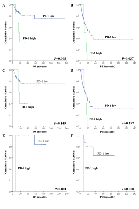
The median follow-up duration for surviving patients was 31.9 (range, 2.4–128.5) months. Sixteen (21.1%) patients received consolidative upfront ASCT after a median of 4 cycles (range 2–4) of HD-MTX-based chemotherapy. Expression of PD-1, PD-L1, and PD-L2 was high in 7.9%, 13.2%, and 42.1% patients, respectively. High PD-1 (HR: 4.95, 95% CI: 1.54–15.86, P=0.007) and MSKCC prognostic scoring (HR: 2.56, 95% CI: 1.17-5.64, P=0.019) were independently associated with inferior overall survival on multivariate analysis. High PD-1 also remained an independent prognostic factor for inferior progression-free survival (HR 2.73, 95% CI: 1.12-6.69, P=0.028), as did MSKCC prognostic scoring (HR: 1.56, 95% CI: 1.09-2.45, P=0.041) on multivariate analysis. However, there were no differences in survival according to the expression levels of PD-L1/PD-L2. Patients with high expression of PD-1 showed significantly shorter survival compared to those with low expression of PD-1 (P=0.008 for OS, and P=0.037 for PFS) (Figure A, B). In a subgroup analysis of 60 patients who did not receive upfront ASCT, high PD-1 expression tended to associate with inferior OS (P=0.145) and PFS (P=0.197) (Figure C, D). However, among 16 patients who received upfront ASCT, high PD-1 expression was significantly associated with inferior OS (P<0.001) and PFS (P=0.008) (Figure E, F).
Conclusion
We found that high PD-1 expression was associated with inferior survival in patients with PCNSL. PD-1 may be considered a biomarker and potential therapeutic target in PCNSL.
Session topic: E-poster
Keyword(s): Non-Hodgkin's lymphoma, Tumor immunology
Type: Eposter Presentation
Background
Programmed cell death 1 (PD-1) and its ligands PD-L1/PD-L2 have been shown to mediate immune evasion in various cancers, but their prognostic roles have not been studied in patients with primary central nervous system lymphoma (PCNSL).
Aims
To evaluate the prognostic role of immunohistochemical PD-1, PD-L1, and PD-L2 expression in immunocompetent patients with PCNSL.
Methods
We performed a retrospective, immunohistochemical study on 76 PCNSL patients at initial diagnosis initially treated homogenously with high-dose methotrexate (HD-MTX)-based chemotherapy, and evaluated the prognostic roles of high PD-1, PD-L1, and PD-L2 expression. The cut-off values for high PD-1 (≥70 cells/high power field [HPF]), PD-L1 (≥100 cells/HPF), and PD-L2 (≥100 cells/HPF) were determined by the area under the receiver operating characteristic curve.
Results
The median follow-up duration for surviving patients was 31.9 (range, 2.4–128.5) months. Sixteen (21.1%) patients received consolidative upfront ASCT after a median of 4 cycles (range 2–4) of HD-MTX-based chemotherapy. Expression of PD-1, PD-L1, and PD-L2 was high in 7.9%, 13.2%, and 42.1% patients, respectively. High PD-1 (HR: 4.95, 95% CI: 1.54–15.86, P=0.007) and MSKCC prognostic scoring (HR: 2.56, 95% CI: 1.17-5.64, P=0.019) were independently associated with inferior overall survival on multivariate analysis. High PD-1 also remained an independent prognostic factor for inferior progression-free survival (HR 2.73, 95% CI: 1.12-6.69, P=0.028), as did MSKCC prognostic scoring (HR: 1.56, 95% CI: 1.09-2.45, P=0.041) on multivariate analysis. However, there were no differences in survival according to the expression levels of PD-L1/PD-L2. Patients with high expression of PD-1 showed significantly shorter survival compared to those with low expression of PD-1 (P=0.008 for OS, and P=0.037 for PFS) (Figure A, B). In a subgroup analysis of 60 patients who did not receive upfront ASCT, high PD-1 expression tended to associate with inferior OS (P=0.145) and PFS (P=0.197) (Figure C, D). However, among 16 patients who received upfront ASCT, high PD-1 expression was significantly associated with inferior OS (P<0.001) and PFS (P=0.008) (Figure E, F).
Conclusion
We found that high PD-1 expression was associated with inferior survival in patients with PCNSL. PD-1 may be considered a biomarker and potential therapeutic target in PCNSL.
Session topic: E-poster
Keyword(s): Non-Hodgkin's lymphoma, Tumor immunology
Abstract: E1383
Type: Eposter Presentation
Background
Programmed cell death 1 (PD-1) and its ligands PD-L1/PD-L2 have been shown to mediate immune evasion in various cancers, but their prognostic roles have not been studied in patients with primary central nervous system lymphoma (PCNSL).
Aims
To evaluate the prognostic role of immunohistochemical PD-1, PD-L1, and PD-L2 expression in immunocompetent patients with PCNSL.
Methods
We performed a retrospective, immunohistochemical study on 76 PCNSL patients at initial diagnosis initially treated homogenously with high-dose methotrexate (HD-MTX)-based chemotherapy, and evaluated the prognostic roles of high PD-1, PD-L1, and PD-L2 expression. The cut-off values for high PD-1 (≥70 cells/high power field [HPF]), PD-L1 (≥100 cells/HPF), and PD-L2 (≥100 cells/HPF) were determined by the area under the receiver operating characteristic curve.
Results
The median follow-up duration for surviving patients was 31.9 (range, 2.4–128.5) months. Sixteen (21.1%) patients received consolidative upfront ASCT after a median of 4 cycles (range 2–4) of HD-MTX-based chemotherapy. Expression of PD-1, PD-L1, and PD-L2 was high in 7.9%, 13.2%, and 42.1% patients, respectively. High PD-1 (HR: 4.95, 95% CI: 1.54–15.86, P=0.007) and MSKCC prognostic scoring (HR: 2.56, 95% CI: 1.17-5.64, P=0.019) were independently associated with inferior overall survival on multivariate analysis. High PD-1 also remained an independent prognostic factor for inferior progression-free survival (HR 2.73, 95% CI: 1.12-6.69, P=0.028), as did MSKCC prognostic scoring (HR: 1.56, 95% CI: 1.09-2.45, P=0.041) on multivariate analysis. However, there were no differences in survival according to the expression levels of PD-L1/PD-L2. Patients with high expression of PD-1 showed significantly shorter survival compared to those with low expression of PD-1 (P=0.008 for OS, and P=0.037 for PFS) (Figure A, B). In a subgroup analysis of 60 patients who did not receive upfront ASCT, high PD-1 expression tended to associate with inferior OS (P=0.145) and PFS (P=0.197) (Figure C, D). However, among 16 patients who received upfront ASCT, high PD-1 expression was significantly associated with inferior OS (P<0.001) and PFS (P=0.008) (Figure E, F).
Conclusion
We found that high PD-1 expression was associated with inferior survival in patients with PCNSL. PD-1 may be considered a biomarker and potential therapeutic target in PCNSL.
Session topic: E-poster
Keyword(s): Non-Hodgkin's lymphoma, Tumor immunology
{{ help_message }}
{{filter}}