ASSESSING MYELOMA BONE DISEASE WITH WHOLE-BODY DIFFUSION-WEIGHTED MRI: COMPARISON WITH FDG PET-CT AND ASSESSMENT OF ADC VALUE
(Abstract release date: 05/19/16)
EHA Library. Narita K. 06/09/16; 132847; E1298

Mr. Kentaro Narita
Contributions
Contributions
Abstract
Abstract: E1298
Type: Eposter Presentation
Background
The recent International Myeloma Working Group (IMWG) consensus statement recommends whole-body magnetic resonance imaging (MRI) for patients with suspected smoldering multiple myeloma (SMM) and solitary plasmacytoma (Dimopoulos et al. 2015). Whole-body diffusion-weighted MRI (WB-DWI) is a promising technique with higher sensitivity for bone lesions and lower cost. However, little is known about its sensitivity compared to 18F-fluorodeoxyglucose positron emission tomography (FDG PET-CT).
Aims
The aim of this study is to compare the sensitivity of myeloma-related bone disease by WB-DWI and FDG PET-CT, and to compare the apparent diffusion coefficients (ADC) of myelomatous lesions with normal healthy controls.
Methods
We identified patients with monoclonal plasma cell disorders that underwent WB-DWI between July 2015 and February 2016 at Kameda Medical Center, Kamogawa-shi, Japan. We compared disease burden by matched simultaneous assessments of WB-DWI and FDG PET-CT using a previously described scoring system based on the number and pattern (focal/diffuse) of disease in each body region (C-spine, T-spine, L-spine, pelvis, ribs/other, long bones). Whole-body score was calculated as the sum of each body lesion (Sharon et al. 2014). Scans performed in patients in different clinical status were excluded. We also analyzed ADC values of WB-DWI in comparison to eight healthy volunteers. Minimal ADC values were measured in each body region (C-spine, T-spine, L-spine, pelvis, ribs/other) and whole-body ADC value was defined as the mean ADC value of each body region. Inversion grayscale maximum intensity projection images of whole-body diffusion-weighted and ACD maps with b value = 900s/mm2 were assessed in this study. All statistical analyses were performed with EZR, which is a graphical user interface for R ver. 3.2.1.
Results
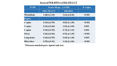
We identified 64 patients that underwent WB-DWI during the study period. Fifty patients underwent FDG PET-CT in the same disease status. Median age of the patients was 71 years (range: 53 – 89) and 23 (44.8%) were male. Three patients had monoclonal gammopathy of undetermined significance (MGUS), five patients had SMM, and 42 patients had symptomatic MM, including five with plasmacytoma. Diffuse lesions were found in 15 (30%) patients by WB-DWI, while FDG PET-CT detected two (4%) diffuse lesions. The detection ratio of focal lesions was the same for both procedures. The whole-body score was significantly higher in WB-DWI than FDG PET-CT (WB-DWI: mean 5.61 ± 8.56 vs. FDG PET-CT: mean 1.08, P < 0.001). In each body region, scores for WB-DWI were significantly higher than those for FDG PET-CT. ADC values were analyzed in 64 patients that underwent diffusion-weighted WB-MRI and compared with eight healthy volunteers. Median age and number of male patients was 71 years (range 47 – 89) and 25 (39.6%), respectively. Four patients were diagnosed as MGUS, eight patients had SMM, and 51 patients had MM, including five with plasmacytoma. The median ADC value of the whole body was significantly lower in patients with monoclonal plasma cell disorders than in the healthy volunteers (680 mm2/s × 10-6 vs. 765.5 mm2/s × 10-6, P = 0.02). In each body region except C-spine and ribs/other, the ADC value was significantly lower in patients with monoclonal plasma cell disorders than in the healthy volunteers.
Conclusion
In patients with monoclonal plasma cell disorders, WB-DWI is more sensitive than FDG PET-CT for detection of bone lesions. Assessment of ADC value is an effective method for detecting bone lesions of monoclonal plasma cell disorders.

Session topic: E-poster
Keyword(s): FDG-PET, Magnetic resonance imaging, Multiple myeloma
Type: Eposter Presentation
Background
The recent International Myeloma Working Group (IMWG) consensus statement recommends whole-body magnetic resonance imaging (MRI) for patients with suspected smoldering multiple myeloma (SMM) and solitary plasmacytoma (Dimopoulos et al. 2015). Whole-body diffusion-weighted MRI (WB-DWI) is a promising technique with higher sensitivity for bone lesions and lower cost. However, little is known about its sensitivity compared to 18F-fluorodeoxyglucose positron emission tomography (FDG PET-CT).
Aims
The aim of this study is to compare the sensitivity of myeloma-related bone disease by WB-DWI and FDG PET-CT, and to compare the apparent diffusion coefficients (ADC) of myelomatous lesions with normal healthy controls.
Methods
We identified patients with monoclonal plasma cell disorders that underwent WB-DWI between July 2015 and February 2016 at Kameda Medical Center, Kamogawa-shi, Japan. We compared disease burden by matched simultaneous assessments of WB-DWI and FDG PET-CT using a previously described scoring system based on the number and pattern (focal/diffuse) of disease in each body region (C-spine, T-spine, L-spine, pelvis, ribs/other, long bones). Whole-body score was calculated as the sum of each body lesion (Sharon et al. 2014). Scans performed in patients in different clinical status were excluded. We also analyzed ADC values of WB-DWI in comparison to eight healthy volunteers. Minimal ADC values were measured in each body region (C-spine, T-spine, L-spine, pelvis, ribs/other) and whole-body ADC value was defined as the mean ADC value of each body region. Inversion grayscale maximum intensity projection images of whole-body diffusion-weighted and ACD maps with b value = 900s/mm2 were assessed in this study. All statistical analyses were performed with EZR, which is a graphical user interface for R ver. 3.2.1.
Results
We identified 64 patients that underwent WB-DWI during the study period. Fifty patients underwent FDG PET-CT in the same disease status. Median age of the patients was 71 years (range: 53 – 89) and 23 (44.8%) were male. Three patients had monoclonal gammopathy of undetermined significance (MGUS), five patients had SMM, and 42 patients had symptomatic MM, including five with plasmacytoma. Diffuse lesions were found in 15 (30%) patients by WB-DWI, while FDG PET-CT detected two (4%) diffuse lesions. The detection ratio of focal lesions was the same for both procedures. The whole-body score was significantly higher in WB-DWI than FDG PET-CT (WB-DWI: mean 5.61 ± 8.56 vs. FDG PET-CT: mean 1.08, P < 0.001). In each body region, scores for WB-DWI were significantly higher than those for FDG PET-CT. ADC values were analyzed in 64 patients that underwent diffusion-weighted WB-MRI and compared with eight healthy volunteers. Median age and number of male patients was 71 years (range 47 – 89) and 25 (39.6%), respectively. Four patients were diagnosed as MGUS, eight patients had SMM, and 51 patients had MM, including five with plasmacytoma. The median ADC value of the whole body was significantly lower in patients with monoclonal plasma cell disorders than in the healthy volunteers (680 mm2/s × 10-6 vs. 765.5 mm2/s × 10-6, P = 0.02). In each body region except C-spine and ribs/other, the ADC value was significantly lower in patients with monoclonal plasma cell disorders than in the healthy volunteers.
Conclusion
In patients with monoclonal plasma cell disorders, WB-DWI is more sensitive than FDG PET-CT for detection of bone lesions. Assessment of ADC value is an effective method for detecting bone lesions of monoclonal plasma cell disorders.

Session topic: E-poster
Keyword(s): FDG-PET, Magnetic resonance imaging, Multiple myeloma
Abstract: E1298
Type: Eposter Presentation
Background
The recent International Myeloma Working Group (IMWG) consensus statement recommends whole-body magnetic resonance imaging (MRI) for patients with suspected smoldering multiple myeloma (SMM) and solitary plasmacytoma (Dimopoulos et al. 2015). Whole-body diffusion-weighted MRI (WB-DWI) is a promising technique with higher sensitivity for bone lesions and lower cost. However, little is known about its sensitivity compared to 18F-fluorodeoxyglucose positron emission tomography (FDG PET-CT).
Aims
The aim of this study is to compare the sensitivity of myeloma-related bone disease by WB-DWI and FDG PET-CT, and to compare the apparent diffusion coefficients (ADC) of myelomatous lesions with normal healthy controls.
Methods
We identified patients with monoclonal plasma cell disorders that underwent WB-DWI between July 2015 and February 2016 at Kameda Medical Center, Kamogawa-shi, Japan. We compared disease burden by matched simultaneous assessments of WB-DWI and FDG PET-CT using a previously described scoring system based on the number and pattern (focal/diffuse) of disease in each body region (C-spine, T-spine, L-spine, pelvis, ribs/other, long bones). Whole-body score was calculated as the sum of each body lesion (Sharon et al. 2014). Scans performed in patients in different clinical status were excluded. We also analyzed ADC values of WB-DWI in comparison to eight healthy volunteers. Minimal ADC values were measured in each body region (C-spine, T-spine, L-spine, pelvis, ribs/other) and whole-body ADC value was defined as the mean ADC value of each body region. Inversion grayscale maximum intensity projection images of whole-body diffusion-weighted and ACD maps with b value = 900s/mm2 were assessed in this study. All statistical analyses were performed with EZR, which is a graphical user interface for R ver. 3.2.1.
Results
We identified 64 patients that underwent WB-DWI during the study period. Fifty patients underwent FDG PET-CT in the same disease status. Median age of the patients was 71 years (range: 53 – 89) and 23 (44.8%) were male. Three patients had monoclonal gammopathy of undetermined significance (MGUS), five patients had SMM, and 42 patients had symptomatic MM, including five with plasmacytoma. Diffuse lesions were found in 15 (30%) patients by WB-DWI, while FDG PET-CT detected two (4%) diffuse lesions. The detection ratio of focal lesions was the same for both procedures. The whole-body score was significantly higher in WB-DWI than FDG PET-CT (WB-DWI: mean 5.61 ± 8.56 vs. FDG PET-CT: mean 1.08, P < 0.001). In each body region, scores for WB-DWI were significantly higher than those for FDG PET-CT. ADC values were analyzed in 64 patients that underwent diffusion-weighted WB-MRI and compared with eight healthy volunteers. Median age and number of male patients was 71 years (range 47 – 89) and 25 (39.6%), respectively. Four patients were diagnosed as MGUS, eight patients had SMM, and 51 patients had MM, including five with plasmacytoma. The median ADC value of the whole body was significantly lower in patients with monoclonal plasma cell disorders than in the healthy volunteers (680 mm2/s × 10-6 vs. 765.5 mm2/s × 10-6, P = 0.02). In each body region except C-spine and ribs/other, the ADC value was significantly lower in patients with monoclonal plasma cell disorders than in the healthy volunteers.
Conclusion
In patients with monoclonal plasma cell disorders, WB-DWI is more sensitive than FDG PET-CT for detection of bone lesions. Assessment of ADC value is an effective method for detecting bone lesions of monoclonal plasma cell disorders.

Session topic: E-poster
Keyword(s): FDG-PET, Magnetic resonance imaging, Multiple myeloma
Type: Eposter Presentation
Background
The recent International Myeloma Working Group (IMWG) consensus statement recommends whole-body magnetic resonance imaging (MRI) for patients with suspected smoldering multiple myeloma (SMM) and solitary plasmacytoma (Dimopoulos et al. 2015). Whole-body diffusion-weighted MRI (WB-DWI) is a promising technique with higher sensitivity for bone lesions and lower cost. However, little is known about its sensitivity compared to 18F-fluorodeoxyglucose positron emission tomography (FDG PET-CT).
Aims
The aim of this study is to compare the sensitivity of myeloma-related bone disease by WB-DWI and FDG PET-CT, and to compare the apparent diffusion coefficients (ADC) of myelomatous lesions with normal healthy controls.
Methods
We identified patients with monoclonal plasma cell disorders that underwent WB-DWI between July 2015 and February 2016 at Kameda Medical Center, Kamogawa-shi, Japan. We compared disease burden by matched simultaneous assessments of WB-DWI and FDG PET-CT using a previously described scoring system based on the number and pattern (focal/diffuse) of disease in each body region (C-spine, T-spine, L-spine, pelvis, ribs/other, long bones). Whole-body score was calculated as the sum of each body lesion (Sharon et al. 2014). Scans performed in patients in different clinical status were excluded. We also analyzed ADC values of WB-DWI in comparison to eight healthy volunteers. Minimal ADC values were measured in each body region (C-spine, T-spine, L-spine, pelvis, ribs/other) and whole-body ADC value was defined as the mean ADC value of each body region. Inversion grayscale maximum intensity projection images of whole-body diffusion-weighted and ACD maps with b value = 900s/mm2 were assessed in this study. All statistical analyses were performed with EZR, which is a graphical user interface for R ver. 3.2.1.
Results
We identified 64 patients that underwent WB-DWI during the study period. Fifty patients underwent FDG PET-CT in the same disease status. Median age of the patients was 71 years (range: 53 – 89) and 23 (44.8%) were male. Three patients had monoclonal gammopathy of undetermined significance (MGUS), five patients had SMM, and 42 patients had symptomatic MM, including five with plasmacytoma. Diffuse lesions were found in 15 (30%) patients by WB-DWI, while FDG PET-CT detected two (4%) diffuse lesions. The detection ratio of focal lesions was the same for both procedures. The whole-body score was significantly higher in WB-DWI than FDG PET-CT (WB-DWI: mean 5.61 ± 8.56 vs. FDG PET-CT: mean 1.08, P < 0.001). In each body region, scores for WB-DWI were significantly higher than those for FDG PET-CT. ADC values were analyzed in 64 patients that underwent diffusion-weighted WB-MRI and compared with eight healthy volunteers. Median age and number of male patients was 71 years (range 47 – 89) and 25 (39.6%), respectively. Four patients were diagnosed as MGUS, eight patients had SMM, and 51 patients had MM, including five with plasmacytoma. The median ADC value of the whole body was significantly lower in patients with monoclonal plasma cell disorders than in the healthy volunteers (680 mm2/s × 10-6 vs. 765.5 mm2/s × 10-6, P = 0.02). In each body region except C-spine and ribs/other, the ADC value was significantly lower in patients with monoclonal plasma cell disorders than in the healthy volunteers.
Conclusion
In patients with monoclonal plasma cell disorders, WB-DWI is more sensitive than FDG PET-CT for detection of bone lesions. Assessment of ADC value is an effective method for detecting bone lesions of monoclonal plasma cell disorders.

Session topic: E-poster
Keyword(s): FDG-PET, Magnetic resonance imaging, Multiple myeloma
{{ help_message }}
{{filter}}


