
11C- METHIONINE PET SCAN FOR THE DETECTION OF MULTIPLE MYELOMA LESIONS
(Abstract release date: 05/19/16)
EHA Library. Figueroa Mora R. 06/09/16; 132818; E1269

Dr. Rocío Figueroa Mora
Contributions
Contributions
Abstract
Abstract: E1269
Type: Eposter Presentation
Background
Multiple Myeloma (MM) is a plasma cell neoplasia that represents the 10-15% of hematological malignancies. The myeloma diagnostic criteria have recently changed based on the availability of more sensitive tools for early detection of the disease. This particularly apply to imaging techniques such as CT, MRI and PET. 11C-methionine is a radiolabeled amino acid that has proven to have higher avidity for protein synthesis. Some recently published works show that 11C-MET PET/TAC is useful for detecting bone and extramedullary lesions in MM and might be a promising tool in the diagnosis and follow up of this disease.
Aims
The purpose of this study is first to evaluate the performance of 11C-MET PET/CT in MM as a diagnostic method compared to 18FDG-PET/CT, and second to analyze the correlation between the metabolic activity and biological parameters of the disease.
Methods
We have reviewed retrospectively the results of 14 patients (50% women, ranged 31-79 years) with MM (12 cases), Smoldering Multiple Myeloma (one case) and solitary plasmacytoma (one case), who simultaneously underwent 11C-MET and 18FDG- PET/CT at our institution. We have analyzed the number and the uptake value (SUVmax) of the lesions observed with both techniques and we have correlated these findings with the biological parameters of the disease (M component, plasma cell percentage in bone marrow, free light chains, immunoglobulins, calcium, renal function). The concordance and correlation between both techniques were analyzed using Cohen’s kappa coefficient and Pearson coefficient (SPPS version 19).
Results
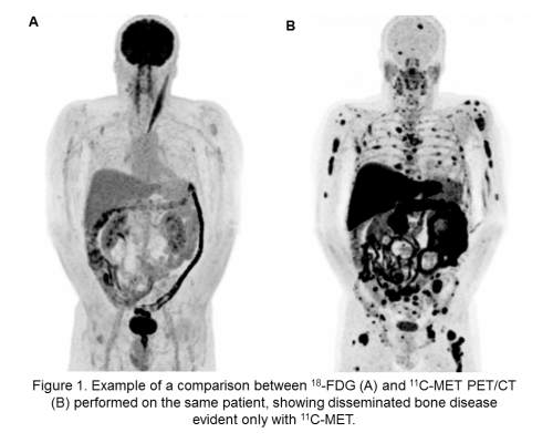
A total of 54 lesions were studied (mean 3.8 lesions/patient). The number of lesions observed with 11C-MET was higher than with 18FDG in 57% of the patients. Only in two cases 18FDG -PET/CT revealed lesions that were not seen with 11C-MET, but interestingly in one of them a biopsy confirmed the non-malignant nature of the lesion. In most cases the SUVmax was higher in 11C-MET for the same lesion compared to 18FDG. Cohen's kappa coefficient between both tests was calculated with a result of 0.29 that means a fair agreement between 18FDG and 11C-MET. This coefficient also reflects a 35% of false negative lesions seen in 18FDG-PET. The following biological parameters were correlated with a significant increase in the uptake value in 11C-MET PET/CT: M-component (r = 0.28; p = 0.02), serum IgG (r = 0.58; p = 0.002), involved free light chain (r = 0.28; p = 0.04) and percentage of plasma cells in the bone marrow (r = 0.32; p = 0.03). These correlations were not significant with the 18FDG PET/CT.
Conclusion
In our experience, PET with 11C- MET could be a highly sensitive tool to evaluate MM lesions. Our data, despite the small number of patients, suggest that it may be superior to conventional 18FDG-PET. Furthermore, there seems to be a significant correlation between some biological parameters, such as FLC, MC or immunoglobulins levels and the avidity of uptake. Larger series are needed to confirm the present findings.
Session topic: E-poster
Keyword(s): Diagnosis, Myeloma, PET
Type: Eposter Presentation
Background
Multiple Myeloma (MM) is a plasma cell neoplasia that represents the 10-15% of hematological malignancies. The myeloma diagnostic criteria have recently changed based on the availability of more sensitive tools for early detection of the disease. This particularly apply to imaging techniques such as CT, MRI and PET. 11C-methionine is a radiolabeled amino acid that has proven to have higher avidity for protein synthesis. Some recently published works show that 11C-MET PET/TAC is useful for detecting bone and extramedullary lesions in MM and might be a promising tool in the diagnosis and follow up of this disease.
Aims
The purpose of this study is first to evaluate the performance of 11C-MET PET/CT in MM as a diagnostic method compared to 18FDG-PET/CT, and second to analyze the correlation between the metabolic activity and biological parameters of the disease.
Methods
We have reviewed retrospectively the results of 14 patients (50% women, ranged 31-79 years) with MM (12 cases), Smoldering Multiple Myeloma (one case) and solitary plasmacytoma (one case), who simultaneously underwent 11C-MET and 18FDG- PET/CT at our institution. We have analyzed the number and the uptake value (SUVmax) of the lesions observed with both techniques and we have correlated these findings with the biological parameters of the disease (M component, plasma cell percentage in bone marrow, free light chains, immunoglobulins, calcium, renal function). The concordance and correlation between both techniques were analyzed using Cohen’s kappa coefficient and Pearson coefficient (SPPS version 19).
Results
A total of 54 lesions were studied (mean 3.8 lesions/patient). The number of lesions observed with 11C-MET was higher than with 18FDG in 57% of the patients. Only in two cases 18FDG -PET/CT revealed lesions that were not seen with 11C-MET, but interestingly in one of them a biopsy confirmed the non-malignant nature of the lesion. In most cases the SUVmax was higher in 11C-MET for the same lesion compared to 18FDG. Cohen's kappa coefficient between both tests was calculated with a result of 0.29 that means a fair agreement between 18FDG and 11C-MET. This coefficient also reflects a 35% of false negative lesions seen in 18FDG-PET. The following biological parameters were correlated with a significant increase in the uptake value in 11C-MET PET/CT: M-component (r = 0.28; p = 0.02), serum IgG (r = 0.58; p = 0.002), involved free light chain (r = 0.28; p = 0.04) and percentage of plasma cells in the bone marrow (r = 0.32; p = 0.03). These correlations were not significant with the 18FDG PET/CT.
Conclusion
In our experience, PET with 11C- MET could be a highly sensitive tool to evaluate MM lesions. Our data, despite the small number of patients, suggest that it may be superior to conventional 18FDG-PET. Furthermore, there seems to be a significant correlation between some biological parameters, such as FLC, MC or immunoglobulins levels and the avidity of uptake. Larger series are needed to confirm the present findings.
Session topic: E-poster
Keyword(s): Diagnosis, Myeloma, PET
Abstract: E1269
Type: Eposter Presentation
Background
Multiple Myeloma (MM) is a plasma cell neoplasia that represents the 10-15% of hematological malignancies. The myeloma diagnostic criteria have recently changed based on the availability of more sensitive tools for early detection of the disease. This particularly apply to imaging techniques such as CT, MRI and PET. 11C-methionine is a radiolabeled amino acid that has proven to have higher avidity for protein synthesis. Some recently published works show that 11C-MET PET/TAC is useful for detecting bone and extramedullary lesions in MM and might be a promising tool in the diagnosis and follow up of this disease.
Aims
The purpose of this study is first to evaluate the performance of 11C-MET PET/CT in MM as a diagnostic method compared to 18FDG-PET/CT, and second to analyze the correlation between the metabolic activity and biological parameters of the disease.
Methods
We have reviewed retrospectively the results of 14 patients (50% women, ranged 31-79 years) with MM (12 cases), Smoldering Multiple Myeloma (one case) and solitary plasmacytoma (one case), who simultaneously underwent 11C-MET and 18FDG- PET/CT at our institution. We have analyzed the number and the uptake value (SUVmax) of the lesions observed with both techniques and we have correlated these findings with the biological parameters of the disease (M component, plasma cell percentage in bone marrow, free light chains, immunoglobulins, calcium, renal function). The concordance and correlation between both techniques were analyzed using Cohen’s kappa coefficient and Pearson coefficient (SPPS version 19).
Results
A total of 54 lesions were studied (mean 3.8 lesions/patient). The number of lesions observed with 11C-MET was higher than with 18FDG in 57% of the patients. Only in two cases 18FDG -PET/CT revealed lesions that were not seen with 11C-MET, but interestingly in one of them a biopsy confirmed the non-malignant nature of the lesion. In most cases the SUVmax was higher in 11C-MET for the same lesion compared to 18FDG. Cohen's kappa coefficient between both tests was calculated with a result of 0.29 that means a fair agreement between 18FDG and 11C-MET. This coefficient also reflects a 35% of false negative lesions seen in 18FDG-PET. The following biological parameters were correlated with a significant increase in the uptake value in 11C-MET PET/CT: M-component (r = 0.28; p = 0.02), serum IgG (r = 0.58; p = 0.002), involved free light chain (r = 0.28; p = 0.04) and percentage of plasma cells in the bone marrow (r = 0.32; p = 0.03). These correlations were not significant with the 18FDG PET/CT.
Conclusion
In our experience, PET with 11C- MET could be a highly sensitive tool to evaluate MM lesions. Our data, despite the small number of patients, suggest that it may be superior to conventional 18FDG-PET. Furthermore, there seems to be a significant correlation between some biological parameters, such as FLC, MC or immunoglobulins levels and the avidity of uptake. Larger series are needed to confirm the present findings.
Session topic: E-poster
Keyword(s): Diagnosis, Myeloma, PET
{{ help_message }}
{{filter}}