
ABNORMAL EXPRESSION OF PYRUVATE KINASE M2 IN MYELOID DENDRITIC CELLS OF THE PATIENTS WITH SEVERE APLASTIC ANEMIA
(Abstract release date: 05/19/16)
EHA Library. Zonghong S. 06/09/16; 132679; E1130

Dr. Shao Zonghong
Contributions
Contributions
Abstract
Abstract: E1130
Type: Eposter Presentation
Background
Severe aplastic anemia (SAA) is a hematologic disease characterized by pancytopenia with severe bone marrow failure. dendritic cells was found to be an antigen-presenting cell (APC) with a most powerful function .Our previous studies have demonstrated that activated myeloid dendritic cells (mDCs) increased in the bone marrow of SAA patients, which could accelerate the polarization of Th1 from Th0 cells, and then initiate the cellular immunity which played an important role in the primary stage of the immune response.
Aims
We further analyzed the protein components of mDC in SAA and explored the possible pathogen that activated mDC.
Methods
Basing on our previous research, we further analyzed the protein components of mDC in SAA as well as normal control, furtherly we applied both FACS western and qRT - PCR technology from a perspective of mRNA and protein to validate our discovery.to explored the possible reason that activated mDC.
Results
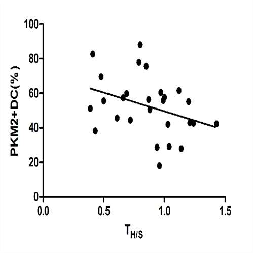
After 7 days of culture, the purity of mDC cells was more than 90% determined by flow cytometry. Results revealed that expression of pyruvate kinase M2(PKM2) were enhanced in mDCs from SAA patients at an early stage of the onset. Concurrently, the level of PKM2 in mDC of SAA patients(59.1±15.8)% was conspicuously higher than that in normal control (32.7±20.2)% at the protein level. We measured the levels of mRNA of PKM2 genes in our patient samples and identified significantly higher expression of PKM2 in the SAA untreated group (1.50±0.84) relative to the recovering group (0.81±0.24) and the control group (0.32±0.11, p<0.05) . In addition, a similar phenomenon was seen by Western blot. As expected, the percentage of PKM2 in mDCs of SAA patients appeared to be strongly correlated with proliferous degree of bone marrow. The percentage of PKM2 in mDCs in patients with SAA was positively correlated with the proportion of reticulocytes absolute neutrophil counts and quantity of mDCs in peripheral blood. Additionally, the ratio of CD4+ T-helper lymphocytes to CD8+ T-suppressor lymphocytes (TH/S) was correlated with the percentage of PKM2 in mDCs. However, no significant difference in platelet counts was detected in SAA patients.Figure legendFigure 1. The purity of mDC cells was more than 90% determined by flow cytometry.Figure 2 . Peptide mass fingerprinting of differentially expressed proteins. After in-gel digestion and mass spectrometry, pyruvate kinase enzyme M2 were expressed higher in the SAA groupFigure 3. Flow cytometry (FCM) tests. The percentage of PKM2 in mDC cells from patients with and normal controls(F3.1), R-SAA(F3.2), severe aplastic anemia(F3.3) are shown.Figure 4. The level of PKM2 in dendritic cell culture supernatant ( pg/ml).Figure 5.The relative PKM2 mRNA level by qPCR.Figure 6.The expression level of PKM2 protein by western blot. Proteins were detected with monoclonal anti-polyhistidine antibodies. We found increased PKM2 expression in untreated SAA patients.Figure 7. Correlation between the proportion of PKM2 in mDCs and clinical parameters in peripheral blood of SAA patients. Data were analyzed using the Spearman correlation coefficient.
Conclusion
These findings demonstrated that dysregulation of PKM2 expression and activation in mDCs might exert an impact on the immune status of SAA patients by enhancing the cellular function of mDC.
Session topic: E-poster
Keyword(s): Dendritic cell, Severe aplastic anemia
Type: Eposter Presentation
Background
Severe aplastic anemia (SAA) is a hematologic disease characterized by pancytopenia with severe bone marrow failure. dendritic cells was found to be an antigen-presenting cell (APC) with a most powerful function .Our previous studies have demonstrated that activated myeloid dendritic cells (mDCs) increased in the bone marrow of SAA patients, which could accelerate the polarization of Th1 from Th0 cells, and then initiate the cellular immunity which played an important role in the primary stage of the immune response.
Aims
We further analyzed the protein components of mDC in SAA and explored the possible pathogen that activated mDC.
Methods
Basing on our previous research, we further analyzed the protein components of mDC in SAA as well as normal control, furtherly we applied both FACS western and qRT - PCR technology from a perspective of mRNA and protein to validate our discovery.to explored the possible reason that activated mDC.
Results
After 7 days of culture, the purity of mDC cells was more than 90% determined by flow cytometry. Results revealed that expression of pyruvate kinase M2(PKM2) were enhanced in mDCs from SAA patients at an early stage of the onset. Concurrently, the level of PKM2 in mDC of SAA patients(59.1±15.8)% was conspicuously higher than that in normal control (32.7±20.2)% at the protein level. We measured the levels of mRNA of PKM2 genes in our patient samples and identified significantly higher expression of PKM2 in the SAA untreated group (1.50±0.84) relative to the recovering group (0.81±0.24) and the control group (0.32±0.11, p<0.05) . In addition, a similar phenomenon was seen by Western blot. As expected, the percentage of PKM2 in mDCs of SAA patients appeared to be strongly correlated with proliferous degree of bone marrow. The percentage of PKM2 in mDCs in patients with SAA was positively correlated with the proportion of reticulocytes absolute neutrophil counts and quantity of mDCs in peripheral blood. Additionally, the ratio of CD4+ T-helper lymphocytes to CD8+ T-suppressor lymphocytes (TH/S) was correlated with the percentage of PKM2 in mDCs. However, no significant difference in platelet counts was detected in SAA patients.Figure legendFigure 1. The purity of mDC cells was more than 90% determined by flow cytometry.Figure 2 . Peptide mass fingerprinting of differentially expressed proteins. After in-gel digestion and mass spectrometry, pyruvate kinase enzyme M2 were expressed higher in the SAA groupFigure 3. Flow cytometry (FCM) tests. The percentage of PKM2 in mDC cells from patients with and normal controls(F3.1), R-SAA(F3.2), severe aplastic anemia(F3.3) are shown.Figure 4. The level of PKM2 in dendritic cell culture supernatant ( pg/ml).Figure 5.The relative PKM2 mRNA level by qPCR.Figure 6.The expression level of PKM2 protein by western blot. Proteins were detected with monoclonal anti-polyhistidine antibodies. We found increased PKM2 expression in untreated SAA patients.Figure 7. Correlation between the proportion of PKM2 in mDCs and clinical parameters in peripheral blood of SAA patients. Data were analyzed using the Spearman correlation coefficient.
Conclusion
These findings demonstrated that dysregulation of PKM2 expression and activation in mDCs might exert an impact on the immune status of SAA patients by enhancing the cellular function of mDC.
Session topic: E-poster
Keyword(s): Dendritic cell, Severe aplastic anemia
Abstract: E1130
Type: Eposter Presentation
Background
Severe aplastic anemia (SAA) is a hematologic disease characterized by pancytopenia with severe bone marrow failure. dendritic cells was found to be an antigen-presenting cell (APC) with a most powerful function .Our previous studies have demonstrated that activated myeloid dendritic cells (mDCs) increased in the bone marrow of SAA patients, which could accelerate the polarization of Th1 from Th0 cells, and then initiate the cellular immunity which played an important role in the primary stage of the immune response.
Aims
We further analyzed the protein components of mDC in SAA and explored the possible pathogen that activated mDC.
Methods
Basing on our previous research, we further analyzed the protein components of mDC in SAA as well as normal control, furtherly we applied both FACS western and qRT - PCR technology from a perspective of mRNA and protein to validate our discovery.to explored the possible reason that activated mDC.
Results
After 7 days of culture, the purity of mDC cells was more than 90% determined by flow cytometry. Results revealed that expression of pyruvate kinase M2(PKM2) were enhanced in mDCs from SAA patients at an early stage of the onset. Concurrently, the level of PKM2 in mDC of SAA patients(59.1±15.8)% was conspicuously higher than that in normal control (32.7±20.2)% at the protein level. We measured the levels of mRNA of PKM2 genes in our patient samples and identified significantly higher expression of PKM2 in the SAA untreated group (1.50±0.84) relative to the recovering group (0.81±0.24) and the control group (0.32±0.11, p<0.05) . In addition, a similar phenomenon was seen by Western blot. As expected, the percentage of PKM2 in mDCs of SAA patients appeared to be strongly correlated with proliferous degree of bone marrow. The percentage of PKM2 in mDCs in patients with SAA was positively correlated with the proportion of reticulocytes absolute neutrophil counts and quantity of mDCs in peripheral blood. Additionally, the ratio of CD4+ T-helper lymphocytes to CD8+ T-suppressor lymphocytes (TH/S) was correlated with the percentage of PKM2 in mDCs. However, no significant difference in platelet counts was detected in SAA patients.Figure legendFigure 1. The purity of mDC cells was more than 90% determined by flow cytometry.Figure 2 . Peptide mass fingerprinting of differentially expressed proteins. After in-gel digestion and mass spectrometry, pyruvate kinase enzyme M2 were expressed higher in the SAA groupFigure 3. Flow cytometry (FCM) tests. The percentage of PKM2 in mDC cells from patients with and normal controls(F3.1), R-SAA(F3.2), severe aplastic anemia(F3.3) are shown.Figure 4. The level of PKM2 in dendritic cell culture supernatant ( pg/ml).Figure 5.The relative PKM2 mRNA level by qPCR.Figure 6.The expression level of PKM2 protein by western blot. Proteins were detected with monoclonal anti-polyhistidine antibodies. We found increased PKM2 expression in untreated SAA patients.Figure 7. Correlation between the proportion of PKM2 in mDCs and clinical parameters in peripheral blood of SAA patients. Data were analyzed using the Spearman correlation coefficient.
Conclusion
These findings demonstrated that dysregulation of PKM2 expression and activation in mDCs might exert an impact on the immune status of SAA patients by enhancing the cellular function of mDC.
Session topic: E-poster
Keyword(s): Dendritic cell, Severe aplastic anemia
{{ help_message }}
{{filter}}